IADR Abstract Archives
Automated Image Analysis For The Quantification Of Oral Dysplasia Severity
Objectives: The current methods for grading oral epithelial dysplasias (OED) are subjective with significant intra- and inter-operator variability. However, the grading guides treatment protocols and influences patient outcomes. Hence, we aimed to develop an automated image analysis tool to objectively quantify differences between the different grades of OED (mild, moderate, severe).
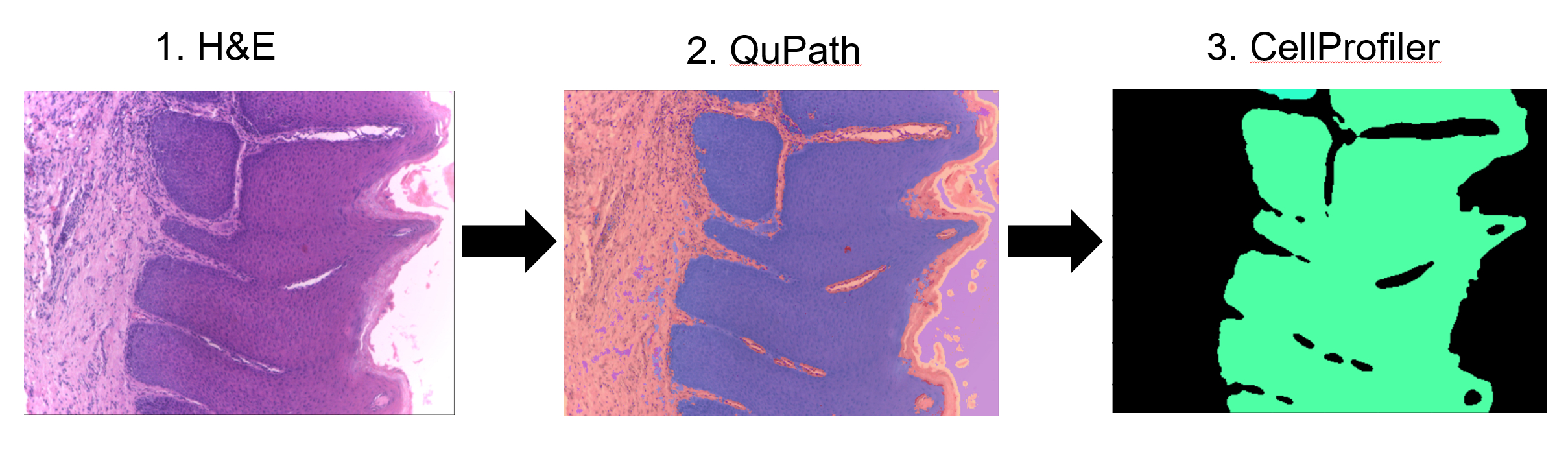
Methods: All H&E-stained photomicrographs of OEDs acquired with a 10x objective were obtained from the publicly available NDB-UFES dataset (n=43). Images of poor quality were excluded (n=6). An image analysis tool was developed using open-source software, QuPath, ImageJ and CellProfiler. A neural network pixel classifier was trained to enable object segmentation of the dysplastic epithelium. Morphometric, textural and granularity measurements are then obtained of the OEDs. Statistical analysis was performed to identify differences between the grades of OEDs.
Results: Quantified features of the OEDs were tested for differences between groups of dysplasia grade by the Wilcoxon rank sum test and clinical features such as age, alcohol and tobacco use were tested with the Fisher’s exact test. Fifteen quantified features were found to be statistically significant (P<0.05). These features related to morphological characteristics of the epithelia such as convex area (P=0.029) and various Zernike features, potentially measuring changes in the architecture of the OEDs. Internal texture (P=0.043) and granularity (P=0.048) features were also significantly different between OED grades and may correspond to measurable cellular changes. None of the clinical features were significantly different between the OED grades.
Conclusions: We have developed an automated image analysis tool for the segmentation and quantification of architectural features of OEDs. We aim to develop the tool further to analyse single-cell features for a more comprehensive, multi-scale analysis. Our preliminary data suggests image features at this scale contain information which may be considered alongside cellular features to aid oral pathologists in the diagnosis and grading of OEDs. This tool has the potential to improve accuracy and reduce intra- and inter-operator variability.
IMAGES
3963898_File000000.jpg
3963898_File000000.jpg