IADR Abstract Archives
Label-free, biomolecular imaging of 3D epithelial tissue equivalents using multiphoton microscopy
Objectives: Drug discovery, tissue engineering and regenerative medicine are entering a new era of biomedical research driven by the demand for three-dimensional (3D) tissue equivalents (TEs) or organoids. Conventional assessment of 3D cultures like histology and confocal microscopy are destructive, time-consuming and/or requires the use of fluorescent tags. Microscopy based on two-photon excitation (TPE) and second harmonic generation (SHG) can be used to address this gap based on their ability to detect endogenous biomolecules like nicotinamide adenine dinucleotide (NAD(P)H), flavin adenine dinucleotide (FAD) and collagen.
Methods: Healthy and hyperproliferative models of 3D epithelial TEs were fabricated using keratinocytes, fibroblast and fibrin-based matrix cultured at air-liquid interface. TPE and SHG imaging were performed using confocal laser scanning microscope equipped with femtosecond pulsed laser excitation source tuned to 740nm for detection of NAD(P)H and 860nm for FAD and collagen. TPE of NAD(P)H and FAD were detected using narrow band pass filters with central wavelengths of 440nm and 628nm respectively, while SHG from collagen detected with 440nm filter.
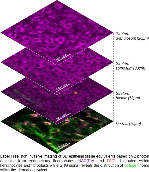
Results: TPE and SHG imaging revealed the different tissue layers of the epithelial TEs. The different layers of keratinocytes (except the corneal layer) strongly expressed NAD(P)H. The cellular size and shape of keratinocytes within individual epithelial layers was distinguishable based on the pattern of perinuclear expression of NAD(P)H. Similarly, fibroblasts expressed both NAD(P)H and FAD enabling the visualization of their orientation in three dimensions. SHG from collagen was clearly visible in the matrix demonstrating the presence of fine fibrils to bundles of collagen fibres. TPE imaging also demonstrated the presence of thicker epithelium in hyperproliferative models.
Conclusions: We demonstrate the potential of multiphoton microscopy for label-free visualization of 3D organization of live, epithelial TEs. Future studies on quantitative biomophologic, biochemical and metabolic features will aid in development of non-invasive tool for clinical and pathophysiological studies.

