IADR Abstract Archives
Spectroscopic evaluation of 3D tissue engineered normal, dysplastic and cancerous oral mucosa
Objectives: Head and neck cancer (HNC) is the sixth most common malignancy worldwide. Squamous cell carcinoma is the primary cause of HNC that evolves from normal epithelium through dysplasia before invading the connective tissue to form a carcinoma. However, less than 18% of doubtful oral lesions proceed to cancer with diagnosis currently relying on histopathological evaluation, which is invasive and time consuming. A non-invasive, real-time, point-of-care method could overcome these problems and facilitate regular screening. Raman spectroscopy can non-invasively generate information regarding the biochemical composition of tissues.
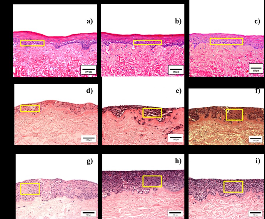
Methods: In this study, Raman Spectroscopy was assessed for its ability to differentiate between normal, pre-malignant and head and neck squamous cell carcinoma (HNSCC). Tissue engineered models of normal, dysplasia and HNSCC were constructed using normal oral keratinocytes, dysplastic and HNSCC cell lines and their biochemical content predicted by interpretation of spectral characteristics.
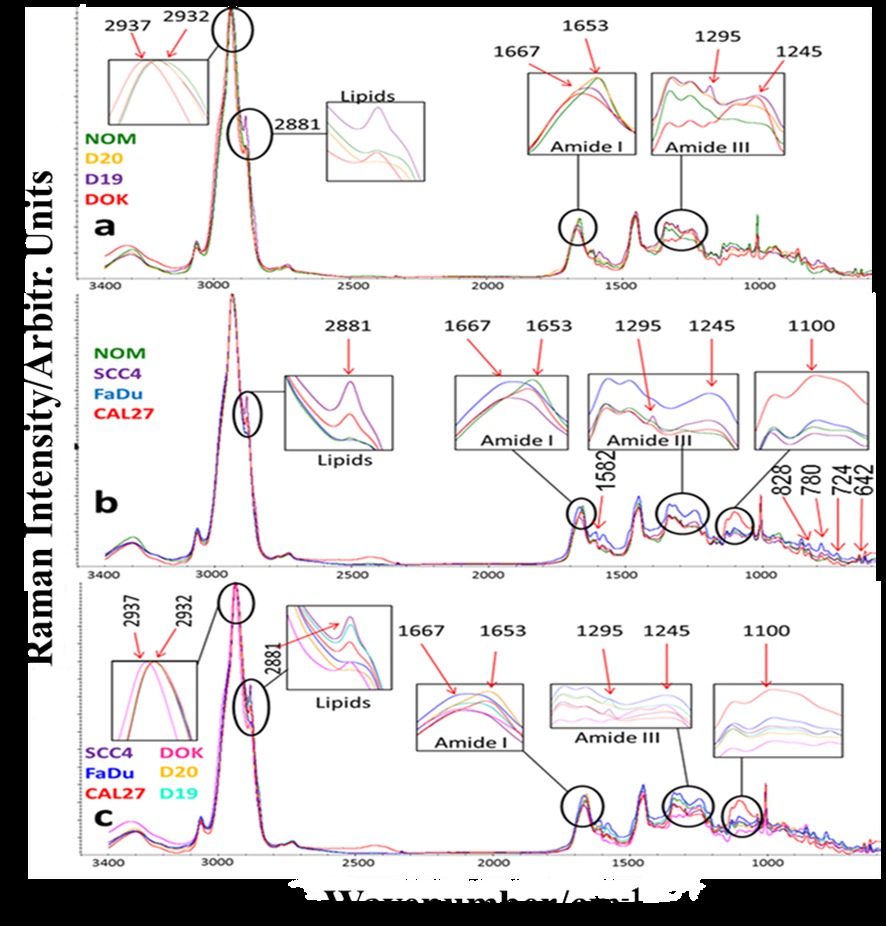
Results: Spectral features of normal models were mainly attributed to lipids, whereas, malignant models were observed to be protein dominant. Visible differences between the spectra of normal, dysplastic and cancerous models, specifically in the bands of amide I and III were observed. Normal mucosal models displayed a sharp and weak lipid peak at 1667cm-1 whereas HNSCC spectra showed a broad and strong amide I peak at this wavenumber. A shift at 2937cm-1 was only observed in DOK, differentiating them from the other tissue types. Multivariate data analysis algorithms successfully identified subtypes of dysplasia and cancer.
Conclusions: 3D Tissue engineered models of oral mucosa provide a valuable platform to investigate complex oral tissues outside the human body. These models closely mimic natural conditions and can be effectively used for applications like therapeutics, diagnostics and treatment purposes. Raman spectroscopy can be used to discriminate between normal and abnormal tissues.
Methods: In this study, Raman Spectroscopy was assessed for its ability to differentiate between normal, pre-malignant and head and neck squamous cell carcinoma (HNSCC). Tissue engineered models of normal, dysplasia and HNSCC were constructed using normal oral keratinocytes, dysplastic and HNSCC cell lines and their biochemical content predicted by interpretation of spectral characteristics.
Results: Spectral features of normal models were mainly attributed to lipids, whereas, malignant models were observed to be protein dominant. Visible differences between the spectra of normal, dysplastic and cancerous models, specifically in the bands of amide I and III were observed. Normal mucosal models displayed a sharp and weak lipid peak at 1667cm-1 whereas HNSCC spectra showed a broad and strong amide I peak at this wavenumber. A shift at 2937cm-1 was only observed in DOK, differentiating them from the other tissue types. Multivariate data analysis algorithms successfully identified subtypes of dysplasia and cancer.
Conclusions: 3D Tissue engineered models of oral mucosa provide a valuable platform to investigate complex oral tissues outside the human body. These models closely mimic natural conditions and can be effectively used for applications like therapeutics, diagnostics and treatment purposes. Raman spectroscopy can be used to discriminate between normal and abnormal tissues.
IMAGES
2360478_File000000.jpg
2360478_File000001.jpg
2360478_File000000.jpg
2360478_File000001.jpg