IADR Abstract Archives
Three-Dimensional Evaluation of Condylar Changes in Female Adolescents With Idiopathic Condylar Resorption Following Stabilization Splint Treatment
Objectives: This study aimed to evaluate the effectiveness of stabilization splint (SS) in treating female adolescents with idiopathic condylar resorption (ICR).
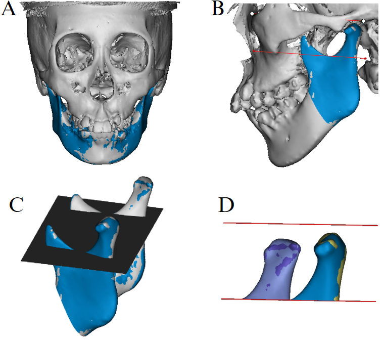
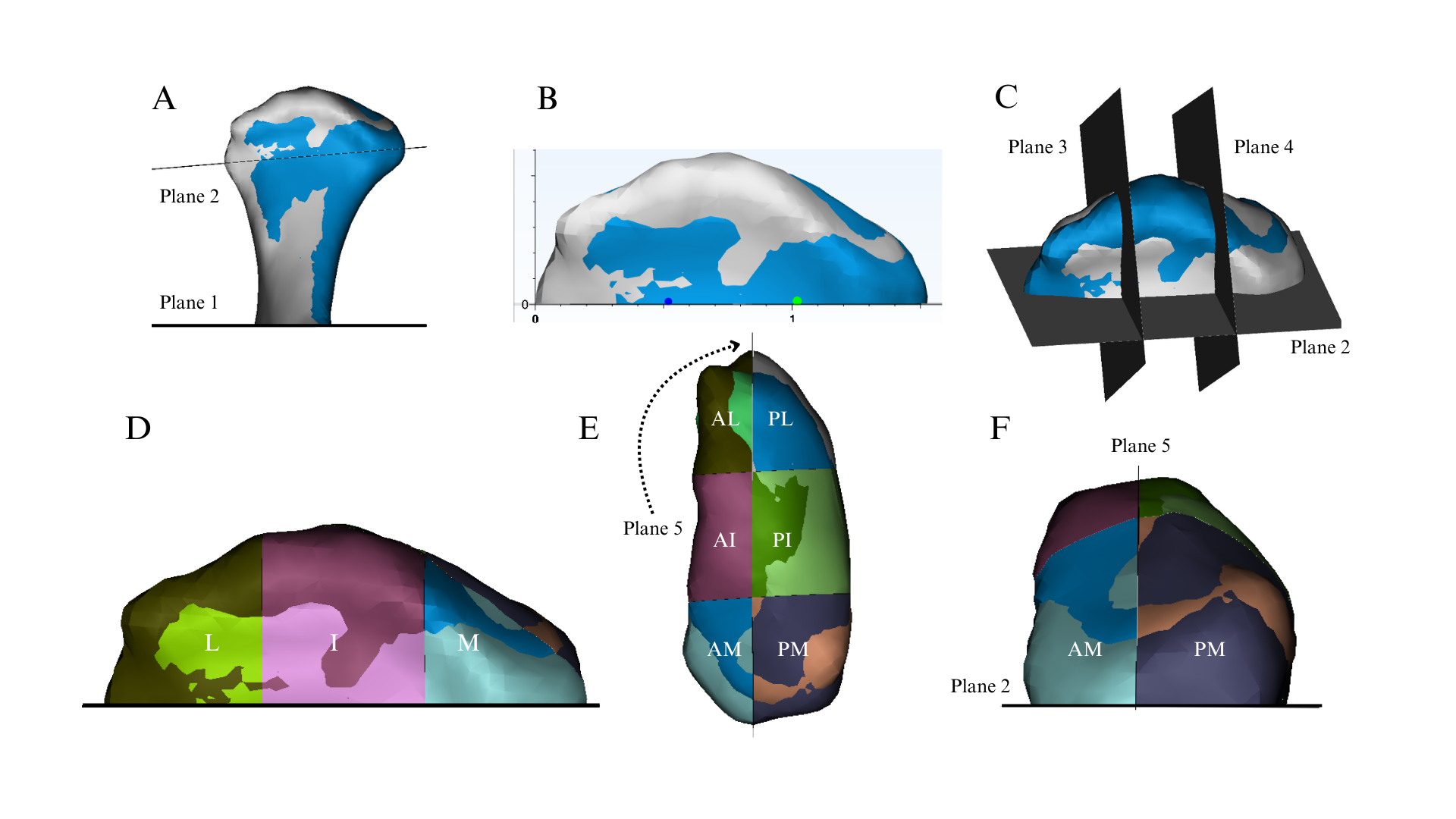
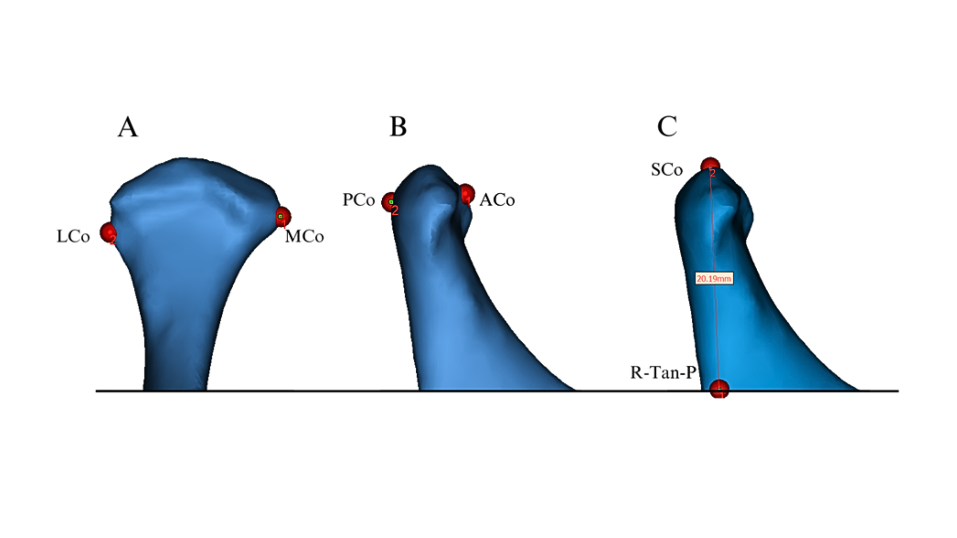
Methods: Thirty-two female patients (mean age: 14.41 ± 2.10 years) diagnosed with ICR were included in this retrospective study. All patients were treated with SS therapy, and cone-beam computed tomography scans were obtained before treatment (T0) and after at least six months with SS treatment (T1), with a mean treatment duration of 7.9±1.97 months. 3D reconstructions assessed the changes in condylar head volume, surface area (SA), and condylar length, width, and height between both time points. To evaluate localized changes, each condylar head was divided into medial, intermediate, and lateral regions, and each region was further divided into anterior and posterior subregions.
Results: Total condylar volume and SA showed no significant differences between T0 and T1. However, regional analysis revealed a significant reduction in volume and SA in the medial region at T1 (mean difference = -7.97 and -9.01, P= 0.011 and 0.004, respectively). The subregional analysis identified significant decreases in volume and SA within the subregions of anterior-medial (AM) (P=0.001 for both) and the anterior-intermediate (AI) (P=0.013 and 0.006, respectively). Condylar height significantly reduced at T1 (MD = -0.25mm, P=0.022), with no significant reduction in condylar width and length.
Conclusions: SS therapy effectively mitigates further condylar resorption and promotes stability in female adolescent ICR patients, particularly by stabilizing overall volume and SA. However, the AM and AI subregions remain susceptible to resorption and require close monitoring.


