IADR Abstract Archives
Temporomandibular Joint Lesions in Patients with Multiple Myeloma
Objectives: The aim of the present study was to evaluate the frequency and pattern of alterations in the temporomandibular joints (TMJ) in patients with multiple myeloma (MM), and to correlate the lesions with the extent of jaw involvement, as well as to correlate the time of their appearance in the course and stage of the disease.
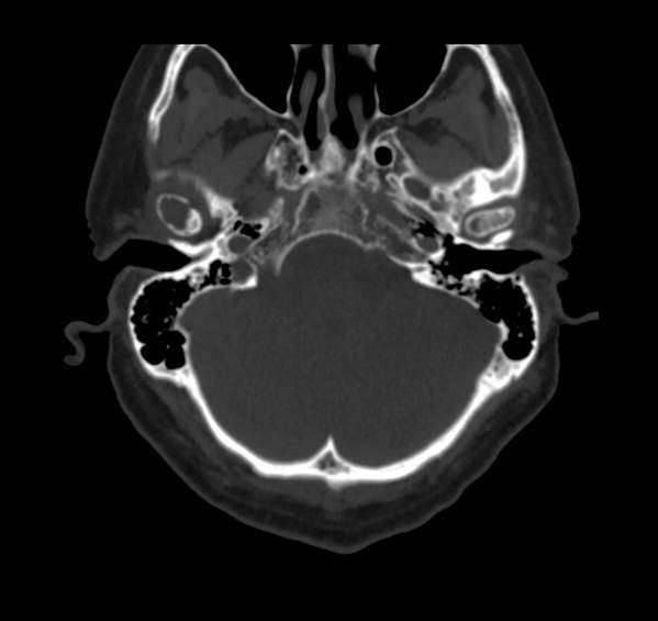
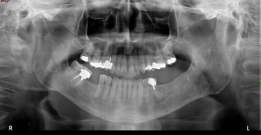
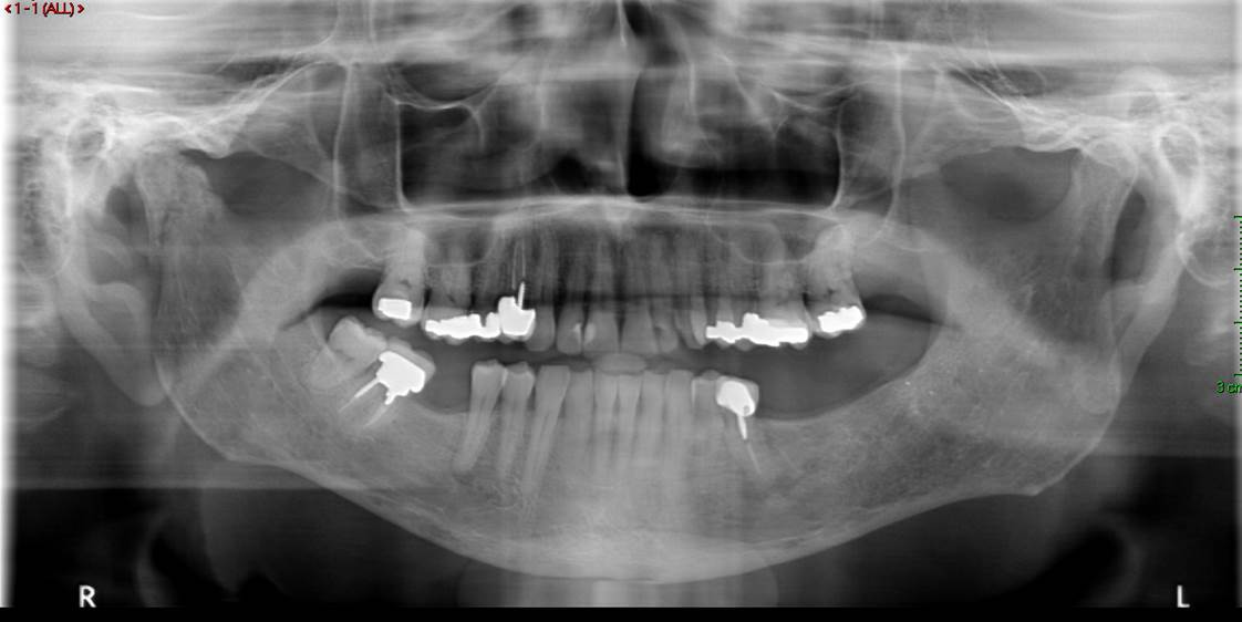
Methods: A total of 122 patients with Multiple Myeloma were evaluated in the outpatient clinic of the department of Oral and Maxillofacial Surgery. All patients had imaging of the jaws in the form of an Orthopantomogram, Computerized Tomography scan, or Magnetic Resonance Imaging. The imaging of the patients was evaluated by a radiologist who specialized in head and neck radiology. Any alteration of the TMJ area that was considered to be myelomatous in origin was documented. Patients with radiological changes in the TMJ were further screened for other lesions of the jaws, and the history and the stage of the Multiple Myeloma were documented carefully.
Results: Twenty-eight patients were found to have myelomatous alterations and lesions of the TMJ. Four of them were symptomatic, and 3 of them presented with pathologic fractures of the mandibular condyle. A thorough description of each patient regarding the duration and stage of MM, previous therapy, and treatment of the TMJ lesion is presented.
Conclusions: The establishment of an accurate diagnosis of a myelomatous lesion in the TMJ of patients with MM can be a diagnostic challenge. The reasons for this include the infrequent occurrence of such lesions, the clinical appearance that mimics a temporomandibular dysfunction syndrome, and the fact that up to half of these lesions are asymptomatic. We recommend a systematic radiologic evaluation of the skeletal system of the jaws (e.g Orthopantomogram) to be performed in subjects with any evidence or suspicion of oral or jaw bone lesions. In addition, any patient with MM that is planned to undergo a dental treatment should undergo a prior orthopantomogram, since punched out lesions carry a risk of a pathologic fracture, occurring spontaneously with no previous trauma. Finally, one must bear in mind, that any of the previously reported cases could have been treated for the TMD complaint without further study for MM involvement, if the routine jaw x-ray examination had not been made.
IMAGES
2290209_File000000.jpg
2290209_File000001.jpg
2290209_File000002.jpg
2290209_File000000.jpg
2290209_File000001.jpg
2290209_File000002.jpg