IADR Abstract Archives
Reparative Calcified Barrier Characterization after Mixing Injectable-Platelet Rich Fibrin with Bioactive Direct Pulp Capping Agents; An Exp. Study.
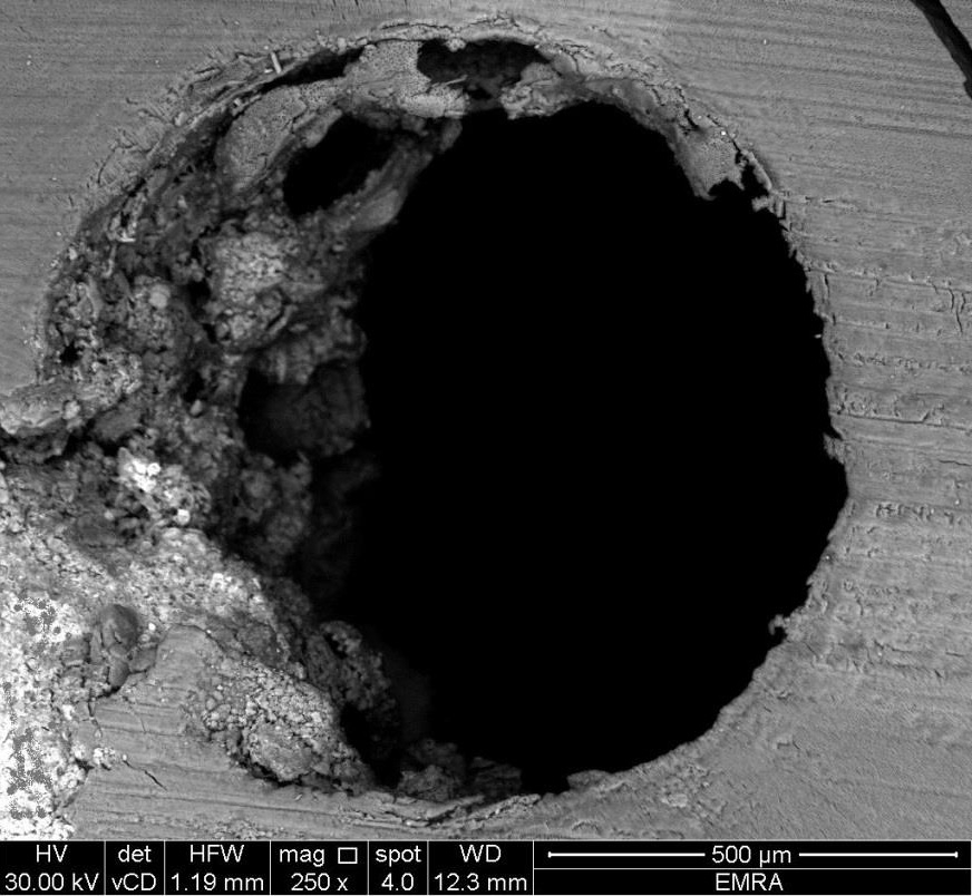
Objectives: This study investigated the morphology and localization of hard tissue barriers formed after direct pulp capping using i-PRF mixed with bioactive agents (Mineral Trioxide Aggregate (MTA) and Bioactive Bone Graft (BBG)) in dogs' teeth using a Scanning Electron Microscope.
Methods: A total of 64 teeth were used out of 8 healthy male mongrel dogs. The teeth were randomly assigned into four groups according to the capping agents used, they were exposed and capped as follows, Group A; capped with MTA, Group B; capped with MTA+ i-PRF, Group C; capped with BBG, Group D; capped with BBG + i-PRF. Finally, the access cavity was restored with Intermediate Restorative Material (IRM). The dogs were euthanized after 1 month, and samples were then prepared for scanning electron microscopic study. SEM was used to assess the morphology, localization, and extension of the reparative hard tissue barriers and use an image-processing and analysis software to delimit the perimeters of the root canal area and the hard tissue barrier to determine the percentage of root canal obliteration.
Results: The chi-square test was used for intragroup comparisons. Results showed that all groups were statistically different (P < 0.05), regarding tissue barrier morphology.
Conclusions: The addition of i-PRF to pulp capping agents allows the production of peripheral hard tissue barriers with more dentinal tubules.
IMAGES
3933161_File000000.jpg
3933161_File000000.jpg