IADR Abstract Archives
Histological Analysis Of Normal And Inflamed Pulp And Stem Cells
Objectives: Evaluate cell morphology of healthy and inflamed human dental pulp and mesenchymal stem cells derived from these tissues.
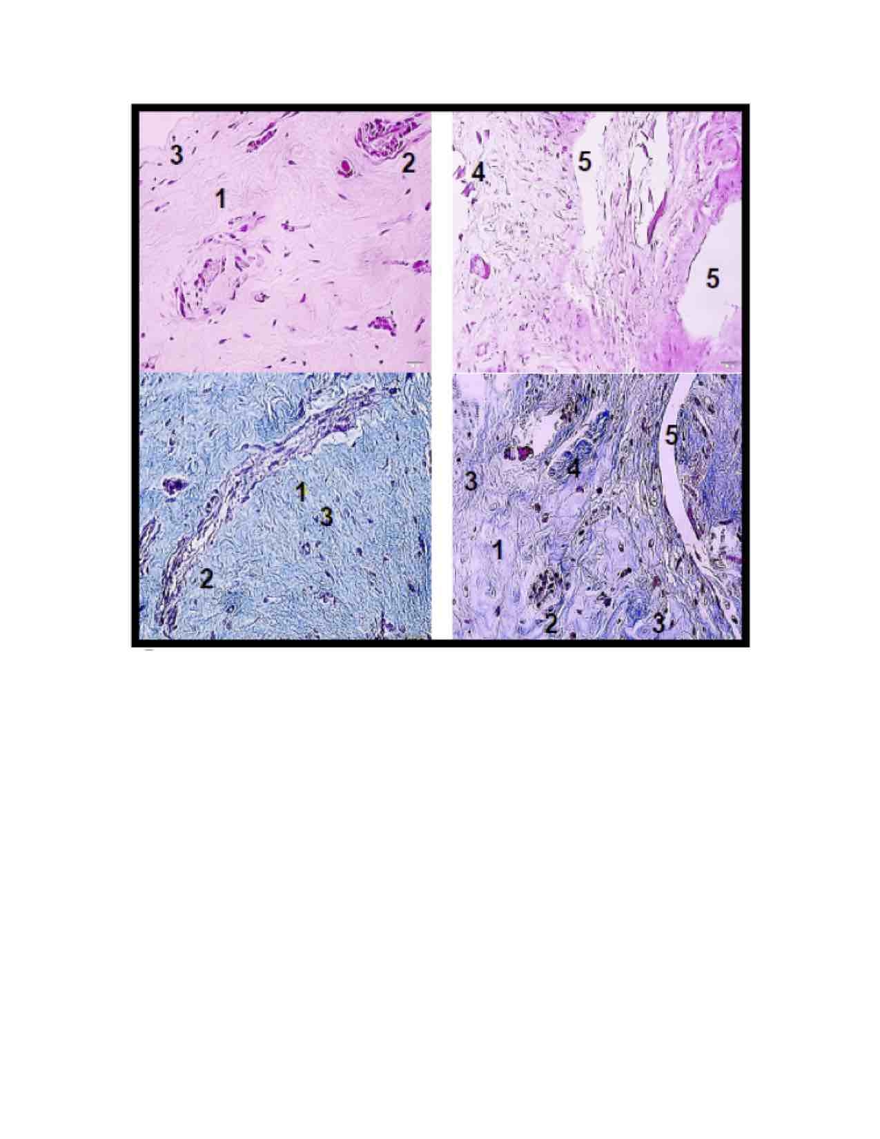
Methods: Dental pulp diagnosed with irreversible pulpitis and normal pulp explants were grown in Alpha-MEM culture medium, 10% SFB, 1% Pen Strep at 37 ° C and 5% CO2. Pulp tissues were fixed in 10% buffered formalin for subsequent histological processing. The mesenchymal stem cells were fixed in 70% ethyl alcohol on 1.9 cm2 culture plates. Subsequently, hematoxylin-eosin and Trichrome Masson's staining of pulp tissues and mesenchymal stem cells were performed. Images were recorded in inverted optical microscopy and images were evaluated by the Image-J program.
Results: There were no differences in cells characteristics and properties such as immunophenotype and tridifferentiation of cells derived from normal and inflamed pulp tissue. The histological dental pulp analysis showed different patterns between healthy and inflamed tissues. The inflamed tissue showed a mostly lax connective tissue and the nuclei were slightly stained. Mesenchymal stem cells derived from both tissues did not show differences in their morphology and phenotypic features typical of a mesenchymal stem cell.
Conclusions: Both pulp tissues showed morphological differences mainly in the lax connective tissue in inflamed dental pulp, however the cells derived from them do not show changes in their morphology
IMAGES
3264886_File000003.jpg
3264886_File000003.jpg