IADR Abstract Archives
Analysis Of Bone Interface In-vivo Following Explantation Using Immersion Endoscopy
Objectives: Quantify by immersion endoscopy the amount of titanium particles adhered to the bone surface at the time of explantation due to periimplantits
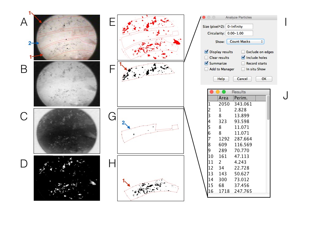
Methods: Explantation by periimplantits and aesthetic compromise in the anterior mandibular area of a bonefit TPS surface implant (Straumann) of a 67-year-old female patient was performed. For the analysis of the bone surface this was divided into a concave and convex portion in relation to the removed implant surface (Figure 1A).The image was processed in 4 steps: B converted in gray, C background inverted, D subtraction of black background with increase of gain and in E threshold appliance. The threshold is a tool that give us the particles without the background. These steps were performed always comparing the TI particles from original figure (A). The regions selected 1 (F and H) and 2 (G) were quantified by the tool Analysis Particles (I) and the amount of TI particles with respective area and perimeter can be analyzed (J). The software used was ImageJ 1.48v.
Results: Statistically significant differences were found in relation to the percentage of Ti particles adhered to the bone surface of explantation with an 18% invasion in the convex areas versus 1% in the concave areas (p <0.005, Independent t test).
Conclusions: The immersion endoscopy technique is a useful clinical tool to identify Ti particles adhering to the bone surface at the time of explantation under high magnification (up to 40 microns). Probably the migration of Ti particles from the surface of the implant to the peri-implant tissues seems to be a little-studied phenomenon that could play an important role in a body-to-stranger reaction over time, depending on the type of treatment surface that the implant possesses.

