IADR Abstract Archives
Digital Histo-Anatomical Dental Analysis: Validation of a non-destructive technique
Objectives: The present work aims to evaluate the accuracy and validate a novel digital dental histo-anatomical analysis method for the study of dental morphology, when compared to chemical enamel dissolution.
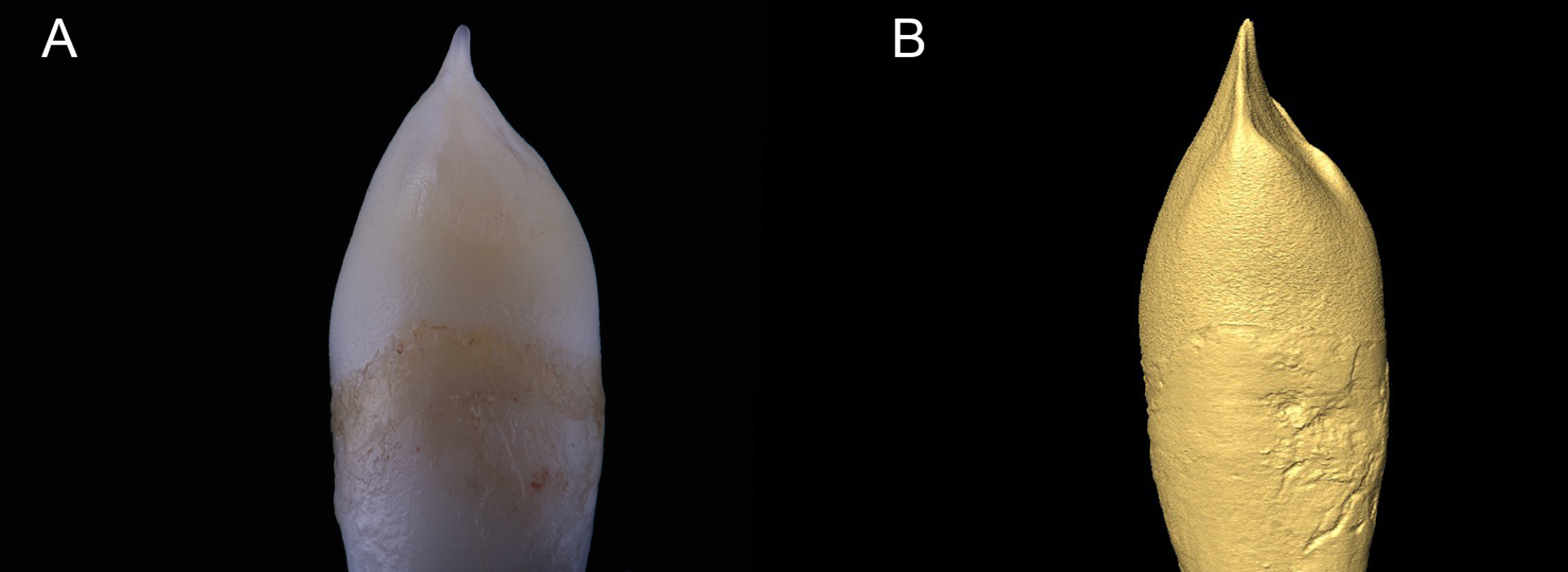
Methods: Extracted maxillary anterior teeth were scanned with micro-computed tomography (μCT 40; Scanco Medical AG), segmented and reconstructed three-dimensionally (Amira v5.5.2). Following digital data acquisition, all specimens were acid-treated with 5% formic acid for careful dissolution of the enamel layer.
Six measurements per specimen were performed, both digitally - following micro-computed tomography scan - and physically – prior and after enamel dissolution. The obtained measurements were subject to statistical analysis through concordance coefficient measurements and linear regression.
Results: A straight line correlation behavior with no statistically significant difference was found between both methods with a concordance correlation coefficient of 97%.
Conclusions: The non-destructive micro-computed tomography layered three-dimensional reconstruction method presented as a reliable non-destructive option for the histo-anatomical analysis of enamel and dentin morphologies.
This study can set a precedent on how dental histo-anatomical studies are performed. Digital three-dimensional data acquisition and rendering methods can provide numerous clinical advantages. Tooth replacement with a diminished sense of loss can be achieved by a rightful combination new technologies with currently available biomaterials.
IMAGES
2809705_File000003.jpg
2809705_File000003.jpg