IADR Abstract Archives
Comparison of Caries Removal Techniques and Dental-Scanners for Quantitative Volume-Loss
Objectives: To evaluate volume loss quantitatively with ICDAS II and also to compare several caries removal techniques and four dental scanners, in terms of volume loss percentages (VLP).
Methods: 120 human molars with ICDAS scores of 3, 4 and 5 were used (n=40). 3D data of each tooth were recorded using an extraoral (Ineos-x5, Dentsply Sirona) and 3 intraoral scanners (iTero-Element 5D (Align), Primescan (Dentsply Sirona), Trios-4 (Trishape) in STL data format. Caries removal techniques were stainless-steel bur (SSB), ceramic bur (CB), tungsten-carbide bur and bioactive-glass powder combination (TCBA), and tungsten-carbide bur and 29µm aluminium-oxide powder combination (TCAl2O3). Following caries removal, secondary STL data were recorded by the scanners. 4 initial and 4 final data for each tooth were overlapped and compared volumetrically using Meshmixer (Autodesk). 960 volumetric data were calculated for 120 teeth. Paired two-sample t test, Wilcoxon test, repeated variance analysis with Bonferroni correction, Friedman test, and Dunn test were used for statistical analyses. Deem significance was set at p<0.050.
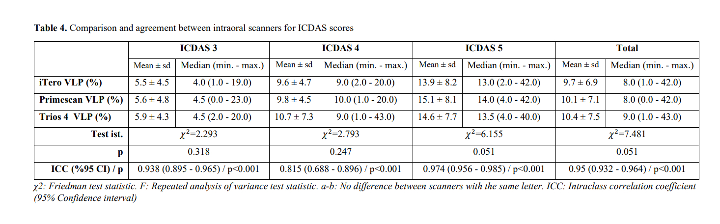
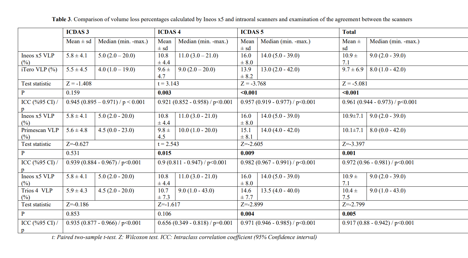
Results: Regarding ICDAS II, VLP for each assessment were considered significant (p<0.001). There was no significant difference in VLP between caries removal techniques (p=0.110). Interactions of caries removal techniques and ICDAS scores were statistically significant (p=0.018). The highest VLP was 18.5% for both SSB and CB groups regarding ICDAS 5. The lowest VLP was 4% for TCBA group regarding ICDAS 3. Significant differences were observed between Ineos-x5 and iTero (p<0.001), Ineos-x5 and Primescan (p=0.001), and Ineos-x5 and Trios-4 (p=0.005). There were no significant differences between iTero, Primescan and Trios-4 (p=0.051).
Conclusions: ICDAS scoring can give preliminary information about volume loss after caries removal procedures. In deep carious lesions, combination of burs and air-abrasion can be useful in preventing substantial tissue loss. The precision of dental scanners can be different regarding the quantitative VLP measurement.
Methods: 120 human molars with ICDAS scores of 3, 4 and 5 were used (n=40). 3D data of each tooth were recorded using an extraoral (Ineos-x5, Dentsply Sirona) and 3 intraoral scanners (iTero-Element 5D (Align), Primescan (Dentsply Sirona), Trios-4 (Trishape) in STL data format. Caries removal techniques were stainless-steel bur (SSB), ceramic bur (CB), tungsten-carbide bur and bioactive-glass powder combination (TCBA), and tungsten-carbide bur and 29µm aluminium-oxide powder combination (TCAl2O3). Following caries removal, secondary STL data were recorded by the scanners. 4 initial and 4 final data for each tooth were overlapped and compared volumetrically using Meshmixer (Autodesk). 960 volumetric data were calculated for 120 teeth. Paired two-sample t test, Wilcoxon test, repeated variance analysis with Bonferroni correction, Friedman test, and Dunn test were used for statistical analyses. Deem significance was set at p<0.050.
Results: Regarding ICDAS II, VLP for each assessment were considered significant (p<0.001). There was no significant difference in VLP between caries removal techniques (p=0.110). Interactions of caries removal techniques and ICDAS scores were statistically significant (p=0.018). The highest VLP was 18.5% for both SSB and CB groups regarding ICDAS 5. The lowest VLP was 4% for TCBA group regarding ICDAS 3. Significant differences were observed between Ineos-x5 and iTero (p<0.001), Ineos-x5 and Primescan (p=0.001), and Ineos-x5 and Trios-4 (p=0.005). There were no significant differences between iTero, Primescan and Trios-4 (p=0.051).
Conclusions: ICDAS scoring can give preliminary information about volume loss after caries removal procedures. In deep carious lesions, combination of burs and air-abrasion can be useful in preventing substantial tissue loss. The precision of dental scanners can be different regarding the quantitative VLP measurement.
IMAGES
3929968_File000000.jpg
3929968_File000001.jpg
3929968_File000000.jpg
3929968_File000001.jpg