IADR Abstract Archives
Remineralization of partially demineralized dentin using calcium-silicate cements
Objectives: To characterize the remineralization potential of calcium-silicate cements (CSC’s) including biomimetic analogs in clinical-like conditions.
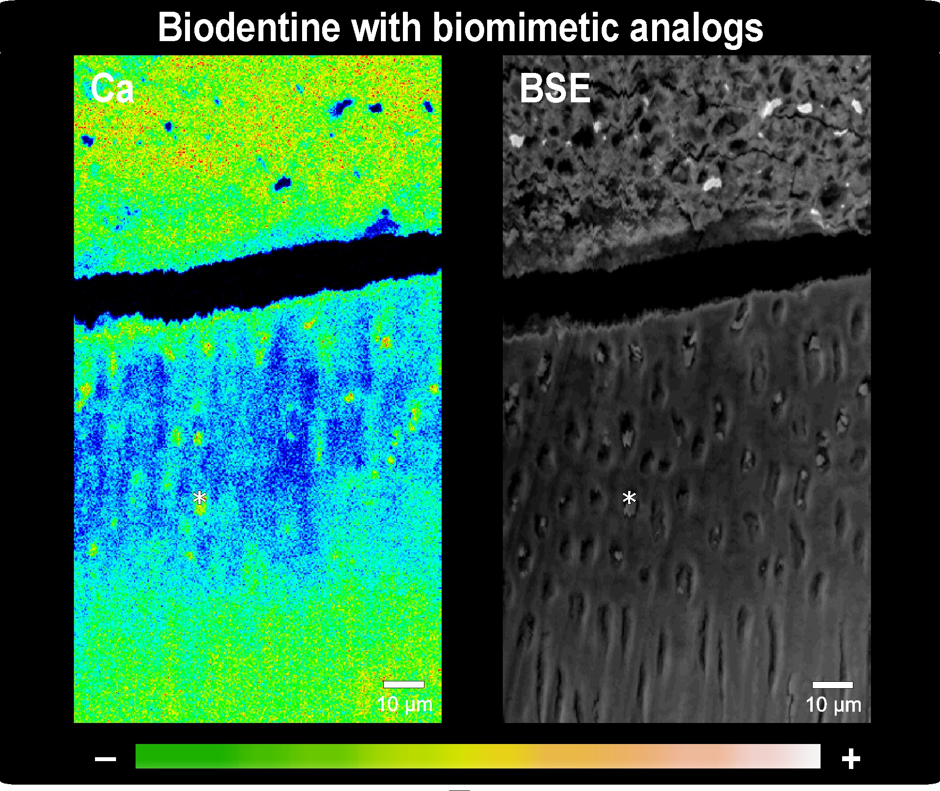
Methods: A class-I dentin cavity was prepared in 24 non-carious third molars. The cavity was coated with a self-etch adhesive (Clearfil SE Bond, Kuraray Noritake), after which a smaller cavity inside was prepared to limit the demineralization area. This area was demineralized using a pH-cycling protocol (50 cyclic immersions in a pH-4.8 and pH-7 bath for 0.5h and 2.5h successively). The cavities were filled with an experimental CSC (‘TCS50’ containing 50% Ca3SiO5 and 50% ZrO2) or the commercial cement Biodentine (Septodont). To half of the specimens, biomimetic analogs (3% polyacrylic acid, 8% sodium tripolyphosphate) were added prior to cement mixing. After 1-week and 6-week storage in phosphate-buffered SBF, the specimens were cross-sectioned and polished using a cross-section polisher (IB-09010CP, JEOL). Interfacial interaction was characterized using a Feg-SEM/EPMA analyzer (JXA-8530F, JEOL).
Results: pH-cycling partially demineralized dentin (100±20 μm thick). After 1-week storage, both cements were found to have filled interfacial voids and to have deposited CaP precipitates within dentin tubules. Also Zr02 was detected 30 μm deep in demineralized dentin in TCS50-filled specimens. No additional interfacial effect by the biomimetic analogs was observed, which may be due to their slow dissolution kinetics. After 6-week storage, tubular occlusion had increased and some indication of initiated intertubular remineralisation was detected at the cement-dentin interface. Adding biomimetic analogs resulted in denser remineralization for both cements. Over time, the cements became more porous, indicating that Ca was transferred to the partially demineralized dentin.
Conclusions: CSC’s with biomimetic analogs were shown to have initiated remineralization of artificial carious-like partially demineralized dentin; 6-week however appeared insufficient to achieve significant remineralization of a 100-μm deep partially demineralized dentin layer. Further in-depth structural investigation of the formed CaP precipitates is also needed.
IMAGES
2300086_File000002.jpg
2300086_File000002.jpg