IADR Abstract Archives
Targeted Biomolecular Expressions at the Mechanically Strained Enthesial Sites of the Dentoalveolar Complex
Objectives: Co-localization of widened and narrowed periodontal spaces (PDL-space) with distribution of nerve bundles (NB), blood vessels (BV), and multi-nucleated cells (MC) proposed to be osteoclasts within a mechanically activated periodontal complex was performed.
Methods: Maxillary molars in 6-week old male C57BL/6 mice (N=5) were experimentally translated for two weeks using an elastic band, with contralateral sides used as internal controls. In situ mechanical testing and imaging using X-ray micro computed tomography (micro-XCT) on the dentoalveolar complex (DC) of a mouse was performed to visualize BV deformation. Widened and narrowed PDL-spaces following tooth movement were correlated with immunolocalized protein gene product 9.5 (PGP 9.5) for light microscopy and osmium tetroxide for nerve distribution in respective groups. Correlative light and electron microscopy (CLEM) images provided nerve distribution within the complex, and NB were further imaged at a higher resolution using scanning transmission electron microscopy (STEM) technique.
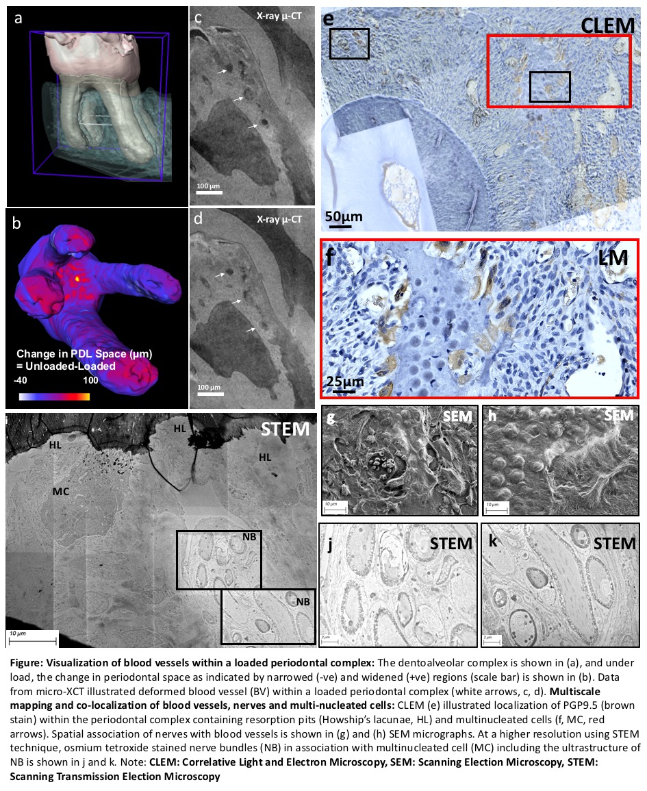
Results: The DC is shown in (fig.a); loading caused changes in PDL-space and are indicated as narrowed (-ve) and widened (+ve) regions (scale bar) (fig.b). Data from micro-XCT illustrated deformed BV within a loaded complex (white arrows, fig.c, fig.d). CLEM image (fig.e) illustrated PGP9.5 localization (brown-DAB stain) adjacent to resorption pits (Howship’s lacunae) and MCs (fig.f, red box, red arrows). Spatial association of nerves with BV is shown in figs. g and h. Osmium tetroxide stained NBs demonstrating ultrastructure and in association with MC are shown in figs.j and h.
Conclusions: Mechanobiological changes in the periodontal complex could involve activation of sensory nerves and vasculature conduction to initiate osteoclastic activity. Mineralization occurs as an attempt to restore PDL-space via remodeling of the PDL, specifically at the entheses. Through extraction of anatomy-specific tissue and associated regenerative molecules using correlative microscopy, this model provides insights into their use as plausible pharmacological agents for tooth movement.
Methods: Maxillary molars in 6-week old male C57BL/6 mice (N=5) were experimentally translated for two weeks using an elastic band, with contralateral sides used as internal controls. In situ mechanical testing and imaging using X-ray micro computed tomography (micro-XCT) on the dentoalveolar complex (DC) of a mouse was performed to visualize BV deformation. Widened and narrowed PDL-spaces following tooth movement were correlated with immunolocalized protein gene product 9.5 (PGP 9.5) for light microscopy and osmium tetroxide for nerve distribution in respective groups. Correlative light and electron microscopy (CLEM) images provided nerve distribution within the complex, and NB were further imaged at a higher resolution using scanning transmission electron microscopy (STEM) technique.
Results: The DC is shown in (fig.a); loading caused changes in PDL-space and are indicated as narrowed (-ve) and widened (+ve) regions (scale bar) (fig.b). Data from micro-XCT illustrated deformed BV within a loaded complex (white arrows, fig.c, fig.d). CLEM image (fig.e) illustrated PGP9.5 localization (brown-DAB stain) adjacent to resorption pits (Howship’s lacunae) and MCs (fig.f, red box, red arrows). Spatial association of nerves with BV is shown in figs. g and h. Osmium tetroxide stained NBs demonstrating ultrastructure and in association with MC are shown in figs.j and h.
Conclusions: Mechanobiological changes in the periodontal complex could involve activation of sensory nerves and vasculature conduction to initiate osteoclastic activity. Mineralization occurs as an attempt to restore PDL-space via remodeling of the PDL, specifically at the entheses. Through extraction of anatomy-specific tissue and associated regenerative molecules using correlative microscopy, this model provides insights into their use as plausible pharmacological agents for tooth movement.
IMAGES
2862990_File000003.jpg
2862990_File000003.jpg