IADR Abstract Archives
Craniofacial deformity and malocclusion in a mouse model of Neurofibromatosis type 1 (NF1)
Objectives: Neurofibromatosis type I (NF 1) is an autosomal dominant disorder affecting approximately 1 in 3500
individuals. NF1 exhibits multiple manifestations such as the presence of café-au-late spots, learning disabilities and
bone deformities. A large proportion of NF 1 patients display skeletal deformities including alteration in bone size and
shape, the presence of scoliosis, and tendency to develop pseudoarthrosis. Craniofacial abnormality occurs in about
7% of NF1 patients and characterized by hypoplasia or absence of greater wing of sphenoid bone. This dysplasia is
progressive and always unilateral, results in bulging of one eye and mid-facial bone associated with malocclusion, and
is termed Sphenoid Wing Dysplasia (SWD). By using an NF1 KO mouse model, we propose to eliminate the
possibility of tumor as a cause of facial deformity, by histological examination of numerous specimens and there by
establish an osteoblast origin for the phenotype, to characterize the time course of the progression of craniofacial
defect and to identify metrics to follow the progression of the phenotype, and to test the effect of treatment with diet,
PTH and Ras antagonist on the development and progression of the phenotype.
Methods: We have established a breeding colony of neurofibromin (NF1 gene) osteoblast conditional knockout mice.
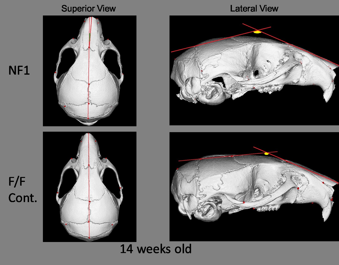
Mice sacrificed for skull Micro-CT scan and for cell harvest ( osteoblast and osteoclast precursors ). The skulls'
images then manipulated and landmarked using Analyze 10.0 software, and the deformity assessed using EDMA
morphometric analysis to compare the changes in landmarks position in control and KO mice skulls.
Results: Results from 12 mice samples ( 8 KO and 4 control) indicate that the NF1ob-/- mice present with cranial
asymmetry with eye bulging and malocclusion as the animal age. Micro-CT of these animals shows a progressive (12-
24 weeks) loss of craniofacial symmetry at the sphenoid bone and other cranial bones. This phenotype is strikingly
similar to SWD seen in NF1 patients.
Conclusions: Upon completion of the study, we will, hopefuly, be able to identify the original cause of sphenoid wing
dysplasia in NF1 and try to block or prevent such deformity, which will help the NF1 patients not to develop
malocclusion and facial asymmetry.

