IADR Abstract Archives
Mixed-Species Biofilms and Artificial Caries Studies of Antibacterial Dental Materials
Objectives: To investigate the effects of experimental antibacterial dental materials (dental composite and bonding agent) on mixed-species biofilms and artificial secondary caries.
Methods: Control composite was formulated with 30wt% dental monomer mixture (BisGMA/EBPADMA/HDDMA= 4:4:2, Esstech, and 1% photoinitiators) and 70wt% UltraFine F-releasing glass filler (SCHOTT AG, Germany). Experimental composites were formulated by replacing part of filler contents with gallium hydroxide loaded mesoporous silica nanoparticles Ga(OH)3@MSN (10wt%) with various amount (3wt%, 4.5wt%, 6 wt%) of synthesized antibacterial monomers C16DC2DMA or BisC11DCDMA. The disk samples (10mm dia, 1.5 mm thick) were light cured and immersed in BHI broth containing 1% sucrose inoculated with four-species consortium, Streptococcus mutans, Lactobacillus casei, S. sanguinis, and Actinomyces naeslundii and incubated at 37°C, 5% CO2 for 24 hours. Biofilms on the disks were removed and plated on BHI agar.
For artificial caries study, class 5 restorations (n=8) were prepared on enamel of extracted teeth and were restored with five groups of materials: Group 1: Control composite and commercial bonding agent (AdperTM ScotchbondTM MP). Group 2 and 3 contained 10wt%Ga(OH)3@MSN plus 4.5wt% of C16DC2DMA and 4.5wt% of BisC11DCDMA, respectively. Group 4 and 5 contained 6wt% C16DC2DMA and BisC11DCDMA. Experimental antibacterial bonding agent was used for Group 2 through 5. After restoration, all teeth were incubated in the above BHI broth for 7 days with medium changed every 48 hours. The specimens were embedded in epoxy resin and sectioned into 0.15mm-thick slices. Images were taken under Nikon i50 polarized light microscope. Wall lesions depth was measured at 50µm distance from adhesive-enamel interface.
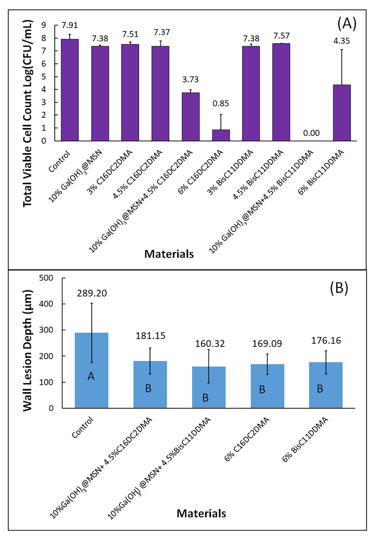
Results: Composites containing 10%Ga(OH)3@MSN and 4.5% antibacterial monomers or 6% antibacterial monomers alone significantly reduced multi-species biofilms (Figure 1A). The same trend is observed in reducing wall lesion depth of secondary caries (Figure 1B).
Conclusions: Experimental antibacterial dental materials can significantly inhibit biofilm and reduce secondary caries.

