IADR Abstract Archives
Root Apex Displacement and Root Resorption (RR) During Orthodontic Treatment
Objectives: Lack of consensus exists regarding the effect of root displacement on RR during orthodontic treatment. Root displacement has been quantified using two-dimensional images. Quantifying treatment-induced root displacement is very difficult using two-dimensional images.
Objectives: To determine the displacement of incisor roots in patients who have received orthodontic treatment with and without extractions.
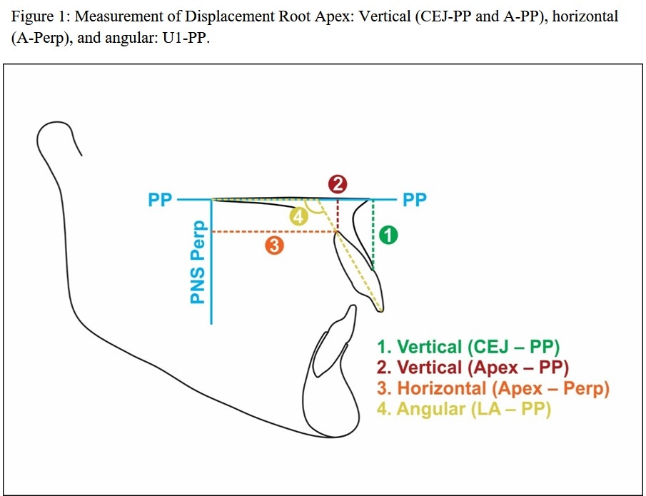
Methods: Records of 215 consecutively treated patients who received orthodontic treatment with or without extractions were obtained for this study. Displacement of maxillary central incisor root was determined using pre- and post-treatment cone beam computerized tomography (CBCT) images using Invivo 5.4. For each patient, either right or left central incisor was randomly chosen to measure the vertical, horizontal and angular displacement of the root (Figure 1) during treatment.
Results: Vertical position of the incisor root as measured by the linear distance between CEJ-PP demonstrated a greater change in the extraction group than non-extraction group. Position of the root apex as measured by Apex-PP distance indicated a greater change in non-extraction group than extraction group (Table1). With similar pre-treatment positions of incisors, the extraction group demonstrated a relatively more tipping compared to the non-extraction group. Change in the angle between long axis of the incisor and palatal plane (PP) during orthodontic treatment demonstrated a large difference between the extraction and non-extraction groups. The extraction group presented with an average pretreatment position of 112.01° with a mean change of -2.54° ± 10.44°. This, in comparison to the non-extraction group which demonstrated a mean change of 8.37°± 7.77°.
Conclusions: Apices of the maxillary incisors moved apically with differing magnitudes and the amount of intrusion in the non-extraction group was more than extraction group. When distance between labial CEJ and PP distance was used an alternative to determine the vertical displacement, roots in the extraction group intruded more than non-extraction group.
Objectives: To determine the displacement of incisor roots in patients who have received orthodontic treatment with and without extractions.
Methods: Records of 215 consecutively treated patients who received orthodontic treatment with or without extractions were obtained for this study. Displacement of maxillary central incisor root was determined using pre- and post-treatment cone beam computerized tomography (CBCT) images using Invivo 5.4. For each patient, either right or left central incisor was randomly chosen to measure the vertical, horizontal and angular displacement of the root (Figure 1) during treatment.
Results: Vertical position of the incisor root as measured by the linear distance between CEJ-PP demonstrated a greater change in the extraction group than non-extraction group. Position of the root apex as measured by Apex-PP distance indicated a greater change in non-extraction group than extraction group (Table1). With similar pre-treatment positions of incisors, the extraction group demonstrated a relatively more tipping compared to the non-extraction group. Change in the angle between long axis of the incisor and palatal plane (PP) during orthodontic treatment demonstrated a large difference between the extraction and non-extraction groups. The extraction group presented with an average pretreatment position of 112.01° with a mean change of -2.54° ± 10.44°. This, in comparison to the non-extraction group which demonstrated a mean change of 8.37°± 7.77°.
Conclusions: Apices of the maxillary incisors moved apically with differing magnitudes and the amount of intrusion in the non-extraction group was more than extraction group. When distance between labial CEJ and PP distance was used an alternative to determine the vertical displacement, roots in the extraction group intruded more than non-extraction group.
3328406_File000000.jpg