IADR Abstract Archives
The Accuracy of Fusion for Intraoral Crown and CBCT Root
Objectives: Obtaining accurate 3-dimensional (3D) crown-root fusion model is critical when developing orthodontic diagnosis and treatment plan.This study’s aim was to develop a new methodology combining intraoral scanning tooth crowns with cone beam CT (CBCT) reconstructing tooth roots for their natural transition in the 3D profile and determine the accuracy of 3D fusion model.
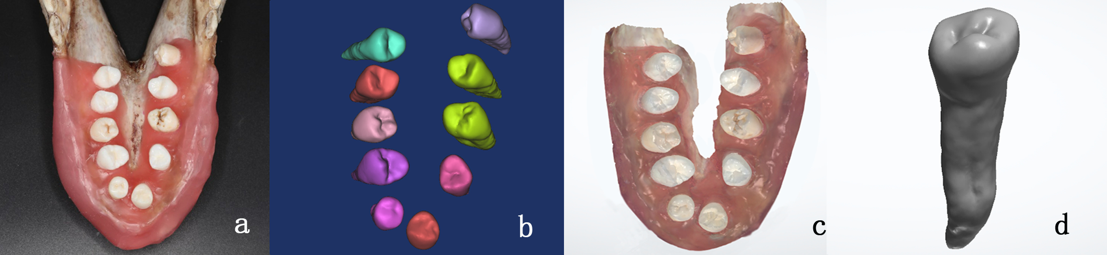
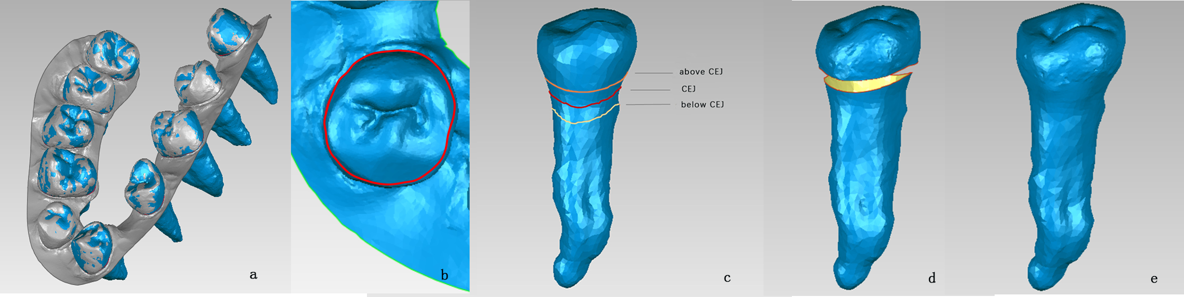
Methods: 3D reference model was established through scanning 10 extracted intact teeth from clinics by the Shining 3D dental scanner . Extracted teeth were then placed in a pig jaw to build a simulated dentition model and scanned by 3-shape intraoral scanner to set up the 3D dentition model. Large field-of-view CBCT imaging was performed with 0.3mm-voxel resolution to construct the dental model with tooth roots .The intraoral dentition model was registered to the CBCT dentition model based on clinical crown in Geomagic Studio 2012.Crown-gingiva boundary was extracted from 3D intraoral dentition model, and then simulated crown-tooth boundary was generated by offsetting and projecting crown-gingiva boundary at different positions. After trimming the crown and root models, the 3D fusion model was formed by curvature or tangent continuity algorithm. Six 3D fusion models were obtained from each tooth ,with 60 fusion models totally. The differences in 3D morphology of intact tooth and anatomic crown between the fusion model and corresponding reference model was calculated using the spectrum colored images and Root Mean Square (RMS) parameter.
Results: A technique to create a fusion model was developed. The accuracy appeared the highest with the method offsetting simulated crown-root boundary above CEJ and curvature continuous algorithm transition ,and mean values of deviation was 0.0813mm in intact tooth,0.0427mm in clinical crown. Analysis of variance showed a statistically significant difference between the positions in offsetting.
Conclusions: This study showed the feasibility of creating a fusion tooth model as well as its accuracy,which could meet the need of orthodontics clinical diagnosis and treatment .The study can provide reference for subsequent research and development automation algorithm programs. However ,the adaption of this method for in vivo model and clinical patients need further research.
IMAGES
3321718_File000000.jpg
3321718_File000001.jpg
3321718_File000000.jpg
3321718_File000001.jpg