IADR Abstract Archives
3D-printed Biodegradable Piezoelectric Scaffold for Bone Regeneration
Objectives: The goal this research was to develop a biodegradable bone regeneration scaffold using poly(L-lactic acid) (PLLA), a biodegradable, electroactive polymer with piezoelectric properties.
Methods: 3D scaffolds were designed and PLLA scaffolds were 3D printed. Scanning electron microscopy(SEM) was used to analyze the surface morphology, and microarchitecture was studied using microCT. Crystallographic characterization was assessed by X-ray diffraction. Compressive modulus was determined using an electromechanical testing system. The piezoelectric potential generated upon mechanical distortion was characterized using an oscilloscope. Hydrolytic degradation was measured as weight loss at internals of 4, 8, and 12 weeks. MG63 cell proliferation was quantified using Cell Counting Kit-8(CCK-8) at 3, 7, 10, and 14 days. Morphology of MG63 cells adhered to the scaffolds was observed using SEM. Cytoskeletal architecture of the cells on the scaffolds was observed using confocal laser scanning microscopy(CLSM) after labelling the cells with Phalloidin-iFluor 488 Reagent and DRAQ5™.
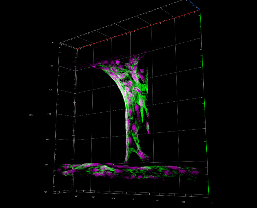
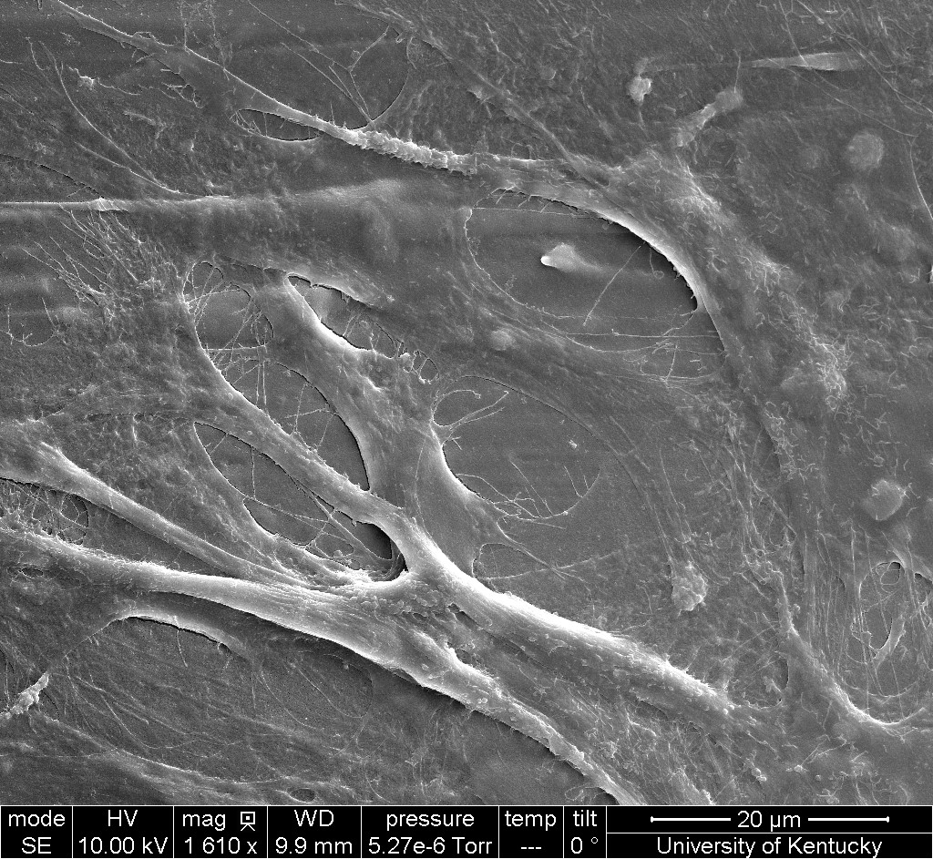
Results: Scaffolds were successfully designed and printed, proving the feasibility of using PLLA for fabricating scaffolds. Average porosity of the scaffold was 53% which is similar to 59% porosity of alveolar trabecular bone. The pore distribution was consistent throughout the scaffold. The average pore size was 197µm, with an average scaffold filament thickness of 183 µm. The average compressive modulus was 204 MPa, which is comparable to 350 MPa of trabecular bone. The crystallinity of polymers was maintained even after the process of melting and 3D printing. The scaffolds exhibited the piezoelectric property by generating electric potential of 50 mV upon cyclic/repeated loading. CCK-8 assay showed cells on scaffold proliferated gradually with time. CLSM(Fig1) and SEM(Fig2) showed cell attachment to the scaffolds.
Conclusions: Porous scaffolds were successfully produced by 3D printing. Test results suggest that the piezoelectric PLLA scaffolds support attachment and proliferation of bone cells and merit further investigation.
Methods: 3D scaffolds were designed and PLLA scaffolds were 3D printed. Scanning electron microscopy(SEM) was used to analyze the surface morphology, and microarchitecture was studied using microCT. Crystallographic characterization was assessed by X-ray diffraction. Compressive modulus was determined using an electromechanical testing system. The piezoelectric potential generated upon mechanical distortion was characterized using an oscilloscope. Hydrolytic degradation was measured as weight loss at internals of 4, 8, and 12 weeks. MG63 cell proliferation was quantified using Cell Counting Kit-8(CCK-8) at 3, 7, 10, and 14 days. Morphology of MG63 cells adhered to the scaffolds was observed using SEM. Cytoskeletal architecture of the cells on the scaffolds was observed using confocal laser scanning microscopy(CLSM) after labelling the cells with Phalloidin-iFluor 488 Reagent and DRAQ5™.
Results: Scaffolds were successfully designed and printed, proving the feasibility of using PLLA for fabricating scaffolds. Average porosity of the scaffold was 53% which is similar to 59% porosity of alveolar trabecular bone. The pore distribution was consistent throughout the scaffold. The average pore size was 197µm, with an average scaffold filament thickness of 183 µm. The average compressive modulus was 204 MPa, which is comparable to 350 MPa of trabecular bone. The crystallinity of polymers was maintained even after the process of melting and 3D printing. The scaffolds exhibited the piezoelectric property by generating electric potential of 50 mV upon cyclic/repeated loading. CCK-8 assay showed cells on scaffold proliferated gradually with time. CLSM(Fig1) and SEM(Fig2) showed cell attachment to the scaffolds.
Conclusions: Porous scaffolds were successfully produced by 3D printing. Test results suggest that the piezoelectric PLLA scaffolds support attachment and proliferation of bone cells and merit further investigation.
IMAGES
3321082_File000000.jpg
3321082_File000001.jpg
3321082_File000000.jpg
3321082_File000001.jpg
