IADR Abstract Archives
Effects of Low Level Laser Therapy in Bone Regeneration of Rabbit Cranial Defects
Objectives: The aim of this study is to determine the levels of bone fill and bone formation with histological, histopathologic and microCT examinations at the 2nd,4th and 8th weeks of rabbit cranial defects of different application protocols of low density (diode) laser. In addition, immunohistochemical evaluations performed at the end of the 2nd week to determine the mechanism of the diode laser.
Methods: 26 New Zealand White Rabbits between 2,5-3,5 kg were used. 8 mm diameter defects were prepared by surgical procedure performed under anesthesia. Three laser doses 4 joules, 6 joules and 8 joules and a control group was utilized. The rabbits received a total of six laser treatments for 10 days, including postoperative application. At the end of the second week, 5 rabbits were sacrificed for histological, immunohistochemical and microCT analyzes. At the 4th and 8th weeks, 20 rabbits were sacrificed for microCT and histopathologic analysis.
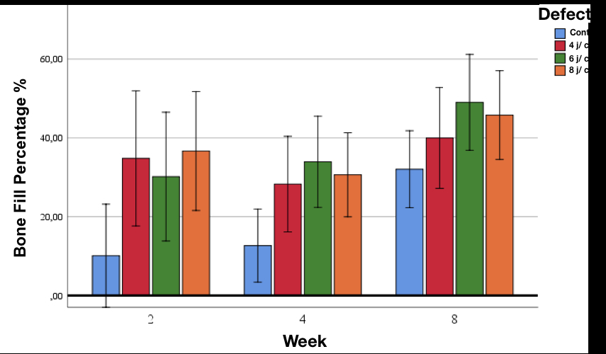
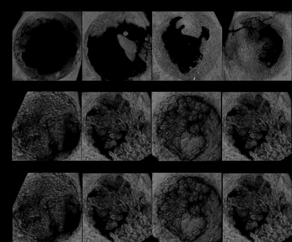
Results: All animals presented favorable healing period and no animals were lost due to surgical approach. Findings of microCT evaluation revealed, more bone fill found in test treated groups compared to non-treated group (p<0.0001) (Fig 1). The most effective laser application group was the 6 joules low intensity laser application group (Fig 2). Histopathological examination revealed more bone islands in the defect wall compared to the control defects. Osteoblastic activity was detected around the bone islands. BMP-2 levels were higher in 4 joule group as shown by immunohistochemical evaluation on sacrificed animals at 2nd weeks. However, this difference is not statistically significant. In osteocalcin staining, osteocalcin expression was significantly higher also in 4 joule group.
Conclusions: In the light of these findings, low-intensity laser application was shown to favor new bone formation in rabbit cranial defects. The high efficacy of doses of 4 j/cm2 and 6 j/cm2 is thought to be promising in applications. Stimulation with diode laser during the early phases of clinical applications such as periodontal regeneration, crest augmentation procedures, and dental implant surgery can be useful to modify and enhance wound healing and regeneration.
Methods: 26 New Zealand White Rabbits between 2,5-3,5 kg were used. 8 mm diameter defects were prepared by surgical procedure performed under anesthesia. Three laser doses 4 joules, 6 joules and 8 joules and a control group was utilized. The rabbits received a total of six laser treatments for 10 days, including postoperative application. At the end of the second week, 5 rabbits were sacrificed for histological, immunohistochemical and microCT analyzes. At the 4th and 8th weeks, 20 rabbits were sacrificed for microCT and histopathologic analysis.
Results: All animals presented favorable healing period and no animals were lost due to surgical approach. Findings of microCT evaluation revealed, more bone fill found in test treated groups compared to non-treated group (p<0.0001) (Fig 1). The most effective laser application group was the 6 joules low intensity laser application group (Fig 2). Histopathological examination revealed more bone islands in the defect wall compared to the control defects. Osteoblastic activity was detected around the bone islands. BMP-2 levels were higher in 4 joule group as shown by immunohistochemical evaluation on sacrificed animals at 2nd weeks. However, this difference is not statistically significant. In osteocalcin staining, osteocalcin expression was significantly higher also in 4 joule group.
Conclusions: In the light of these findings, low-intensity laser application was shown to favor new bone formation in rabbit cranial defects. The high efficacy of doses of 4 j/cm2 and 6 j/cm2 is thought to be promising in applications. Stimulation with diode laser during the early phases of clinical applications such as periodontal regeneration, crest augmentation procedures, and dental implant surgery can be useful to modify and enhance wound healing and regeneration.
IMAGES
3309381_File000001.jpg
3309381_File000002.jpg
3309381_File000001.jpg
3309381_File000002.jpg