IADR Abstract Archives
Cone-Beam Computed Tomography in Osteogenesis Imperfecta: Three-Dimensional Evaluation of Craniofacial Features
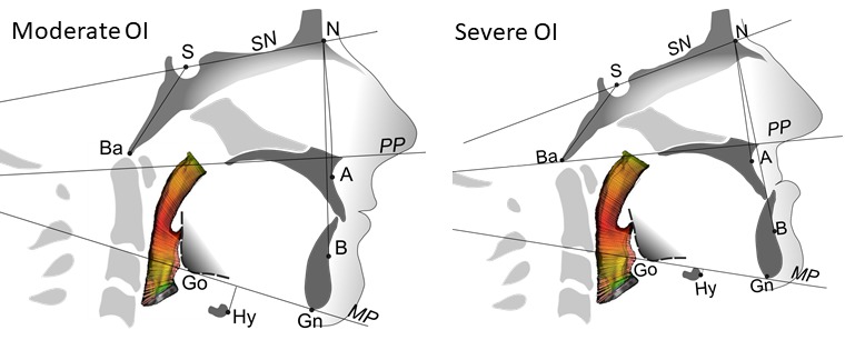
Objectives: Osteogenesis Imperfecta (OI) is a genetic disorder that is predominantly characterized by fragile bones. Apart from fractures, deformities of long bones, scoliosis and stunted growth are hallmarks of the more severe forms of OI. Together with overt craniofacial abnormalities, the skeletal and dental manifestations are highly heterogeneous and have not been investigated with three-dimensional imaging. In the present study, we have used cone-beam computed tomography to assess in 3D craniofacial features and upper airway morphology in moderate to severe OI.
Methods: Cone-beam computed tomography was performed in 41 individuals (age from 10-35 years; 28 females) with moderate to severe OI (OI type III or IV). Angular and linear craniofacial measurements, facial proportions, and airway volume were determined and compared to reference ranges based on literature values for non-OI controls.
Results: Many of the abnormal craniofacial features in OI were associated with an increased cranial base angle. The maxilla was retrognathic and was a significant contributor to the observed malocclusion. The total face height was significantly reduced due to counter-clockwise rotation of the mandible. Both maxilla and mandible were narrower, and together with an increased bi-temporal width, produced the characteristic triangular face appearance. Face asymmetry was common, and cranial asymmetry could be severe. Craniofacial deformities were not associated with reduced patency of the upper airways. Height z-score had a limited association with the craniofacial manifestations of OI.
Conclusions: OI affects the development of the entire craniofacial complex. The cranial base angle reflects the severity of craniofacial abnormalities in OI. A hypoplastic maxilla is a significant contributor to the malocclusion. Despite reports of frequent sleep apnea in OI, we found normal airway morphology.
IMAGES
2957331_File000000.jpg
2957331_File000000.jpg