IADR Abstract Archives
A Comparative Evaluation of Two 3D Printing Hydroxyapatite/Collagen Materials and Their Osteogenic Effects on BMSCs
Objectives: To produce two kinds of 3D printing scaffolds with different morphology of HA, Nano hydroxyapatite (nHA) and deproteinized bovine bone (DBB) dispersed into Collagen (CoL) respectively and to compare the osteogenic effect on human bone mesenchyme stem cells (BMSCs).
Methods: 3D printing scaffolds were produced by the 3D-Plotter (Envision Tec, German). The X-ray photoelectron spectroscopy (XPS) were used to examine the chemical element of the two HA crystals. The Fourier transform infrared spectroscopy (FTIR) and X-ray diffraction (XRD) were used to detect the chemical bonds and crystal phases of the HA combined with Collagen. The Scanning Electron Microscope (SEM) were used to examine the surface morphology of the two kinds of the scaffolds.
BMSCs were cultured on the two groups of 3D printed scaffolds and one control group. After culturing for 1, 3, 5, and7 days, cell proliferation was determined using the CCK-8 method. Immunofluorescent staining were used to observe the cells adhered to the scaffolds under Confocal Laser Scanning Microscopy (CLSM). Real-time quantitative polymerase chain reaction (RT-PCR) was performed to analyze the expression of osteogenesis-related genes (RUNX2, SOX9, OCN and CoL1A1).
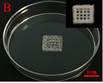
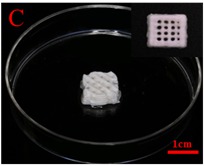
Results: nHA/CoL and DBB/CoL 3D printing scaffolds were showed in images respectively. The composition analysis of XPS, FTIR and XRD indicated that the HA crystals were consistent in the elements but different in the chemical bonds and crystal phases. The SEM results exhibited the different surface morphology of the nHA and DBB scaffolds.
There were no significant difference between the two scaffolds in the cell proliferation. Immunofluorescent staining indicated BMSCs can adhere to the 3D printed structure very well. In vitro osteogenic induction results showed that the expression levels of RUNX2, SOX9, OCN and CoL1A1 were significantly increased after days 7 compared with the control group (p<0.05).
Conclusions: 3D printing scaffolds consisting of nHA/CoL and DBB/CoL both are promising candidate for the individual customized bone substitutes in the future.
Methods: 3D printing scaffolds were produced by the 3D-Plotter (Envision Tec, German). The X-ray photoelectron spectroscopy (XPS) were used to examine the chemical element of the two HA crystals. The Fourier transform infrared spectroscopy (FTIR) and X-ray diffraction (XRD) were used to detect the chemical bonds and crystal phases of the HA combined with Collagen. The Scanning Electron Microscope (SEM) were used to examine the surface morphology of the two kinds of the scaffolds.
BMSCs were cultured on the two groups of 3D printed scaffolds and one control group. After culturing for 1, 3, 5, and7 days, cell proliferation was determined using the CCK-8 method. Immunofluorescent staining were used to observe the cells adhered to the scaffolds under Confocal Laser Scanning Microscopy (CLSM). Real-time quantitative polymerase chain reaction (RT-PCR) was performed to analyze the expression of osteogenesis-related genes (RUNX2, SOX9, OCN and CoL1A1).
Results: nHA/CoL and DBB/CoL 3D printing scaffolds were showed in images respectively. The composition analysis of XPS, FTIR and XRD indicated that the HA crystals were consistent in the elements but different in the chemical bonds and crystal phases. The SEM results exhibited the different surface morphology of the nHA and DBB scaffolds.
There were no significant difference between the two scaffolds in the cell proliferation. Immunofluorescent staining indicated BMSCs can adhere to the 3D printed structure very well. In vitro osteogenic induction results showed that the expression levels of RUNX2, SOX9, OCN and CoL1A1 were significantly increased after days 7 compared with the control group (p<0.05).
Conclusions: 3D printing scaffolds consisting of nHA/CoL and DBB/CoL both are promising candidate for the individual customized bone substitutes in the future.
IMAGES
2954875_File000000.jpg
2954875_File000001.jpg
2954875_File000000.jpg
2954875_File000001.jpg