IADR Abstract Archives
Stress Analysis of Ultrasonic Tip Vibration on Root Canal Wall
Objectives: This study aimed to establish a three-dimensional finite-element model of ultrasonic tip vibration at a fixed point on a root canal wall and analyze the level and distribution of stress in the root canal wall at the start of vibration.
Methods: An extracted mandibular first premolar was chosen for sample preparation, and its intrinsic frequency was measured using a laser vibrometer. The tooth sample was scanned by micro-computed tomography. Then, using the ANSYS software, a finite-element model was established, and its intrinsic frequency was computed and compared with experimental data for validation. The validated finite-element model was used to simulate the vibration of ultrasonic tip ET20 at 5 mm below the root canal orifice at intermediate power (level 5) and perform stress analysis.
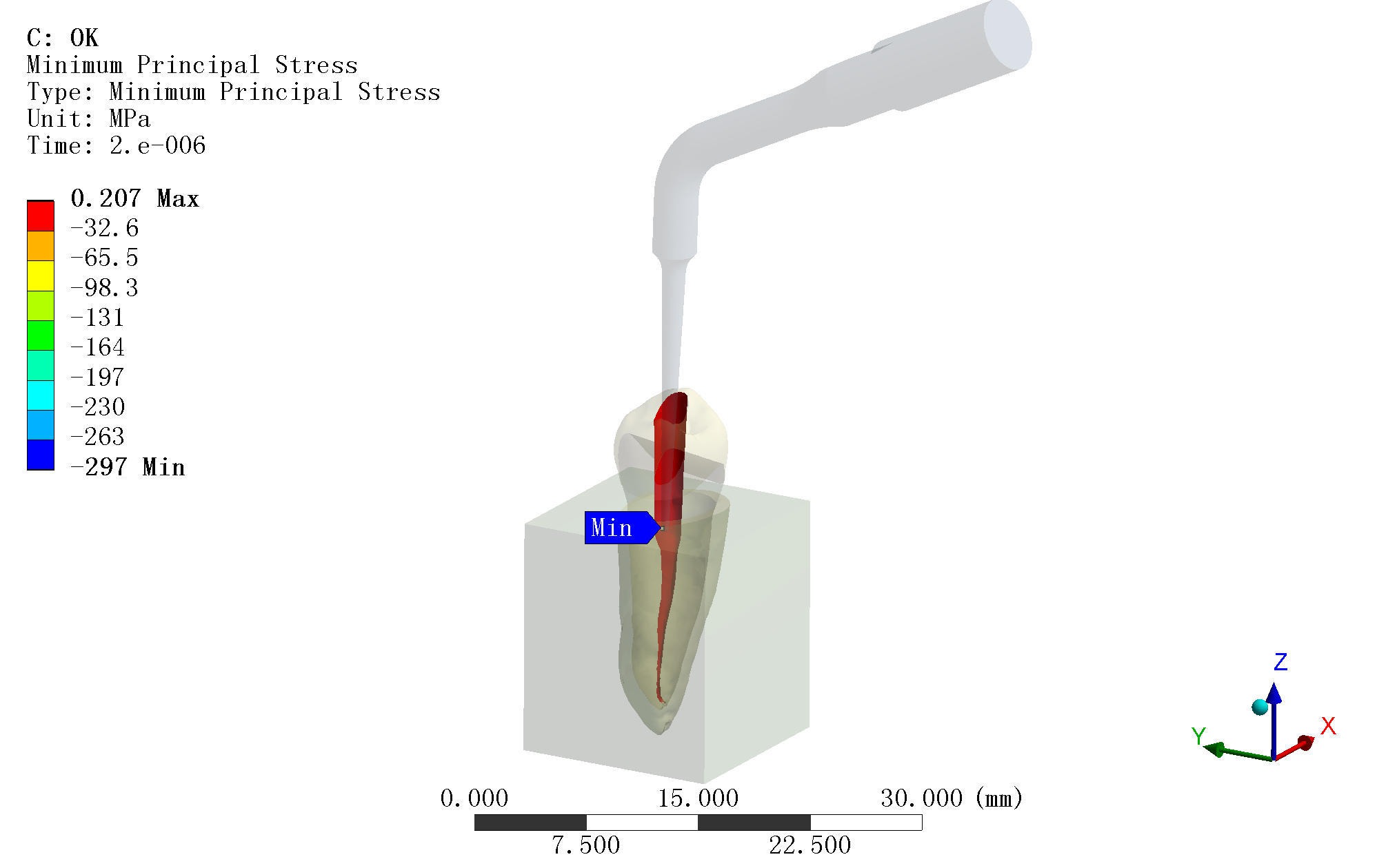
Results: The experimental data of intrinsic frequency of the sample and the results of finite-element computation were in agreement, thus validating the finite-element model. When the E20 tip vibrated at the root canal wall with intermediate power (level 5), the third principal stress at the contact point reduced sharply, instantly reaching the ultimate compressive strength (UCS) of -297 MPa, while the first principal stress was 13.49 MPa — far less than the ultimate tensile strength (UTS). Stress values at the root canal wall outside the contact point was very low — far less than the UCS and UTS.
Conclusions: The three-dimensional finite-element model of ultrasonic tip vibration at a fixed point on a root canal wall exhibits high precision and could be used in future studies on ultrasonic instrumentation. Upon vibration of the ET20 tip at the root canal wall with intermediate power (level 5), the compressive stress at the contact point instantly reached the UCS, which would have subsequently caused dentine compressive failure.
Methods: An extracted mandibular first premolar was chosen for sample preparation, and its intrinsic frequency was measured using a laser vibrometer. The tooth sample was scanned by micro-computed tomography. Then, using the ANSYS software, a finite-element model was established, and its intrinsic frequency was computed and compared with experimental data for validation. The validated finite-element model was used to simulate the vibration of ultrasonic tip ET20 at 5 mm below the root canal orifice at intermediate power (level 5) and perform stress analysis.
Results: The experimental data of intrinsic frequency of the sample and the results of finite-element computation were in agreement, thus validating the finite-element model. When the E20 tip vibrated at the root canal wall with intermediate power (level 5), the third principal stress at the contact point reduced sharply, instantly reaching the ultimate compressive strength (UCS) of -297 MPa, while the first principal stress was 13.49 MPa — far less than the ultimate tensile strength (UTS). Stress values at the root canal wall outside the contact point was very low — far less than the UCS and UTS.
Conclusions: The three-dimensional finite-element model of ultrasonic tip vibration at a fixed point on a root canal wall exhibits high precision and could be used in future studies on ultrasonic instrumentation. Upon vibration of the ET20 tip at the root canal wall with intermediate power (level 5), the compressive stress at the contact point instantly reached the UCS, which would have subsequently caused dentine compressive failure.
IMAGES

2954565_File000000.jpg
2954565_File000001.jpg
2954565_File000000.jpg
2954565_File000001.jpg