IADR Abstract Archives
Facial Shape Analysis in Osteogenesis Imperfecta Using Automated Computer Vision
Objectives:
1. Determine differences in facial morphology between individuals with osteogenesis imperfecta (OI) and controls.
2. Compare the facial phenotype between OI types.
Methods:
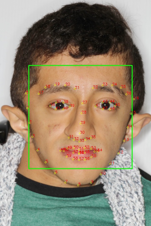
Anteroposterior facial photographs of 184 individuals with OI and 100 healthy control patients with a normal facial index of 0.80 were processed using a python script. This software automatically placed 68 facial landmarks and compiled their 2D coordinates (see image).
The R programming language was used to conduct a statistical shape analysis. Mean shapes were compared using Goodall’s F-Test. Landmark distances were computed using the control mean shape as a baseline. These values were compiled as a mean landmark distance for each patient and were used to determine the group Z-score for each OI type.
Three facial ratios were also computed as follows:
Distances of the lateral edges of the temples to:
1. Distance of lateral edges of the eyes.
2. Angles of the mandible.
3. The height of the face.
Results: The Goodall F-test confirmed that facial form differed between OI and control subjects (p < 0.05). Comparison between OI types I, III and IV revealed that the facial shape of OI type III is different from OI types I and IV (p < 0.05), but no difference was found between OI types I and IV.
The three facial ratios showed statistically significant differences of each OI type from control and intergroup except between for type I and IV.
The group Z-Score for the distances from the control group was of 1.015 for Type I, 2.404 for Type III and 1.279 for Type IV. Notice that type III has a greater Z-score indicating that the facial morphology of patients from this group is significantly different from controls as well as type I and IV patients.
Conclusions: OI is associated with specific facial characteristics such as an increased width of the temples in relation to the rest of the face. This analysis reveals that severity of the facial alterations depend on the OI type, and are most severe in OI type III.
The similarities between the facial shapes of type I and IV subjects challenge our current classification of the facial manifestations of the disease.

