IADR Abstract Archives
Tooth Axis Calculation From CT Images
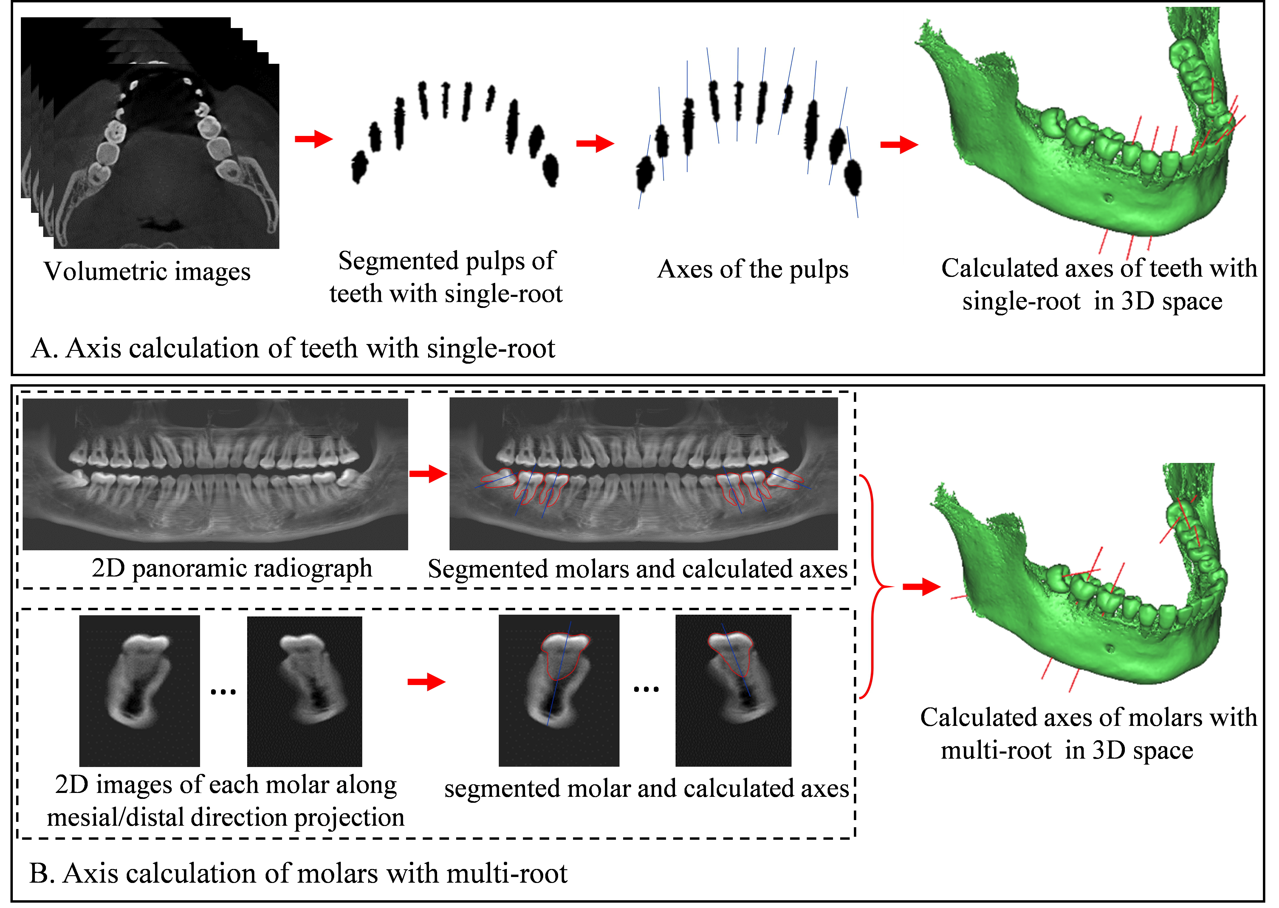
Objectives: The tooth axis is needed for the navigation of dental implant. Currently, it is mainly determined by dentists manually, which is subjective and tedious. This study aimed to develop an automatic method for the calculation of tooth axis from dental CT images.
Methods: To calculate the axes of teeth with single-root, the three-dimensional (3D) dental pulps are segmented from the volumetric CT images using region growing algorithm. The corresponding axes are then calculated from the pulp region using the principal components analysis (PCA). To calculate the axes of molars with multi-root whose pulp boundaries are not clear in the branches of roots, a two-dimensional (2D) panoramic radiograph is synthesized by projecting the volumetric images along the perpendicular direction of the dental arch. Each 2D molar region is then extracted from the panoramic radiograph using region growing algorithm, and 2D axes in the panoramic radiograph are calculated from the extracted region by combing PCA and symmetry axes detection. For each molar, a 2D projection image is synthesized by projecting 3D local images along the mesial-distal direction. 2D axes in the planes of the projection images are then calculated using the same method as in the panoramic radiograph. The corresponding two 2D axes of each molar are used to obtain a 3D molar axis. CT images of 10 subjects were used to test the developed method. Three dentists were required to qualitatively judge whether each calculated tooth axis is accurate enough for the application of dental implant.
Results: All the calculated tooth axes were judged to be accurate enough for the application of dental implant.
Conclusions: The presented axis calculation methods for single-root tooth and multi-root molar are effective to extract the tooth axis from dental CT images.
IMAGES
2476302_File000000.jpg
2476302_File000000.jpg