IADR Abstract Archives
The Biological Properties of Dental Implant Surafce Modified With Novel Technique
Objectives: Dental implant has been very popular nowadays. Previous research pointed out that implant surface treatment may play an important role on osseointegration. The most commonly used dental implant materials are commercially pure titanium and Ti6Al4V. In natural environment, these titanium alloy mantle a cover of titanium dioxide passive layer in short time. Nevertheless, the cover of titanium dioxide is amorphous type and its depth is about 5-6 nm. The titanium dioxide passive layer is helpful on osseointegration, but not stable, and easily broken.
For producing a more intensive and thicker cover of titanium dioxide to provide better environment for osteoblastic cells, atomic layer deposition (ALD) technique is used. In this research, we tested different thickness(control and 100 nm)and two different crystal forms(amorphous, anatase)of titanium dioxide coating using ALD technique. The hypothesis is that thicker titanium dioxides passive layer as expected to be more biocompatible. Scanning electron microscope were carried out in material analyses. Cell viability and F-actin stain were carried out in cell experiments.
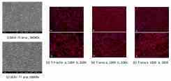
Methods: The titanium (Ti) discs, 8mm in diamter and 1 mm in hight, were fabricated, and polished with grit number 4000 sandpapers. The discs were cleaned and divided into three goups: control (without coating, Ti), amorphous TiO2 ALD coating 100 nm (Ti amo), and anatase TiO2 ALD coating 100 nm (Ti ana). The morphology of discs was observed with SEM. To evalute the biocompalibility, HEPM (human embryonic palatal mesenchyme stem cell ) cell line was cultured for 7 days and used for MTT assay and F-actin stain. The ANOVA and post-hoc Tukey test were used. (p<0.05)
Results: The morphology of amorphous TiO2 ALD coating presented large particle with polygon or oval shape. Ti ana 100 nm showed elongated grain structure with close packing. The growth of HEPM cells was better in Ti amo and Ti ana than Ti, with statististic significant. The F-actin stain revealed that (1) the cells of Ti amo and Ti ana groups was more numerous then Ti group, which proved MTT results (2) the morphology of Ti amo and Ti ana groups showed spindle with more and elonged processes from cell membrane.
Conclusions: The modified amorphous and anatase TiO2 coating with ALD technique improved cell growth and good biocompatibility.
For producing a more intensive and thicker cover of titanium dioxide to provide better environment for osteoblastic cells, atomic layer deposition (ALD) technique is used. In this research, we tested different thickness(control and 100 nm)and two different crystal forms(amorphous, anatase)of titanium dioxide coating using ALD technique. The hypothesis is that thicker titanium dioxides passive layer as expected to be more biocompatible. Scanning electron microscope were carried out in material analyses. Cell viability and F-actin stain were carried out in cell experiments.
Methods: The titanium (Ti) discs, 8mm in diamter and 1 mm in hight, were fabricated, and polished with grit number 4000 sandpapers. The discs were cleaned and divided into three goups: control (without coating, Ti), amorphous TiO2 ALD coating 100 nm (Ti amo), and anatase TiO2 ALD coating 100 nm (Ti ana). The morphology of discs was observed with SEM. To evalute the biocompalibility, HEPM (human embryonic palatal mesenchyme stem cell ) cell line was cultured for 7 days and used for MTT assay and F-actin stain. The ANOVA and post-hoc Tukey test were used. (p<0.05)
Results: The morphology of amorphous TiO2 ALD coating presented large particle with polygon or oval shape. Ti ana 100 nm showed elongated grain structure with close packing. The growth of HEPM cells was better in Ti amo and Ti ana than Ti, with statististic significant. The F-actin stain revealed that (1) the cells of Ti amo and Ti ana groups was more numerous then Ti group, which proved MTT results (2) the morphology of Ti amo and Ti ana groups showed spindle with more and elonged processes from cell membrane.
Conclusions: The modified amorphous and anatase TiO2 coating with ALD technique improved cell growth and good biocompatibility.
IMAGES
2474924_File000022.jpg
2474924_File000022.jpg