IADR Abstract Archives
Analysis of White Spot Lesions Using Scanning Electron Microscopy
Objectives: A white spot lesion (WSL) is a subsurface enamel porosity from carious demineralisation on the smooth surfaces of the tooth with a white opacity appearance. Lesions show an intact surface layer, followed underneath by the more porous lesion body. The small pores within the body of the lesion act as a diffusion pathway for both acids and minerals, so allowing the demineralisation of enamel to occur at the advancing front of the lesion.
The objective is to characterize the porosity structure and its size on WSL with the novel scanning electron microscopy.
Methods: A one micron thick layer of platinum over 25μmx 25μm of the interest region of enamel was deposited by FIB-SEM. Then, a rough cut (25μmx 5μmx 20μm) with 3nA current and 30Kv was applied with the help of drift suppression (DS), using a standard “cross-sectional” cutting pattern, which ended at the front of the deposited platinum layer. Two adjacent areas (25μmx 5μmx 20μm) on the both sides of the platinum layer were milled under the same conditions. Subsequent, the sub-surface edge of interest perpendicular to the surface was polished. The "slice and view" was carried out for milling 1000 slices with 30Kv and 0.5nA and backscattered (BS) images were taken with 30Kv and 4pA. Images were imported into imageJ and analyzed.
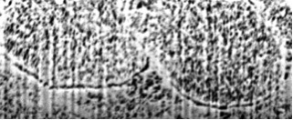
Results: The WSL shows dissolution of prism boundaries as well as internal porosity within the prism itself on FIB-SEM. The porosity size is in a wide range between 100- 500nm.
Conclusions: FIB-SEM is useful to characterize the porosity structure and size of WSL. WSLs show similar structure in the different areas oof WSL with wide range of porosity size.
The objective is to characterize the porosity structure and its size on WSL with the novel scanning electron microscopy.
Methods: A one micron thick layer of platinum over 25μmx 25μm of the interest region of enamel was deposited by FIB-SEM. Then, a rough cut (25μmx 5μmx 20μm) with 3nA current and 30Kv was applied with the help of drift suppression (DS), using a standard “cross-sectional” cutting pattern, which ended at the front of the deposited platinum layer. Two adjacent areas (25μmx 5μmx 20μm) on the both sides of the platinum layer were milled under the same conditions. Subsequent, the sub-surface edge of interest perpendicular to the surface was polished. The "slice and view" was carried out for milling 1000 slices with 30Kv and 0.5nA and backscattered (BS) images were taken with 30Kv and 4pA. Images were imported into imageJ and analyzed.
Results: The WSL shows dissolution of prism boundaries as well as internal porosity within the prism itself on FIB-SEM. The porosity size is in a wide range between 100- 500nm.
Conclusions: FIB-SEM is useful to characterize the porosity structure and size of WSL. WSLs show similar structure in the different areas oof WSL with wide range of porosity size.
2474658_File000000.jpg