IADR Abstract Archives
A Fourier Transform Approach to Modeling Kinetics of Appositional Growth in Healthy vs. Diseased Mineralized Tissues
Objectives: This study investigates shifts in mineral apposition rate (MAR) and growth linearity factor (RW) in bone, dentin, and cementum of periodontally diseased fibrous joints compared to age-matched controls using the principles of Fourier transforms.
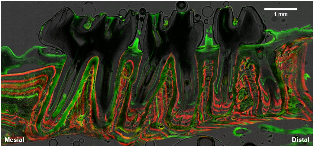
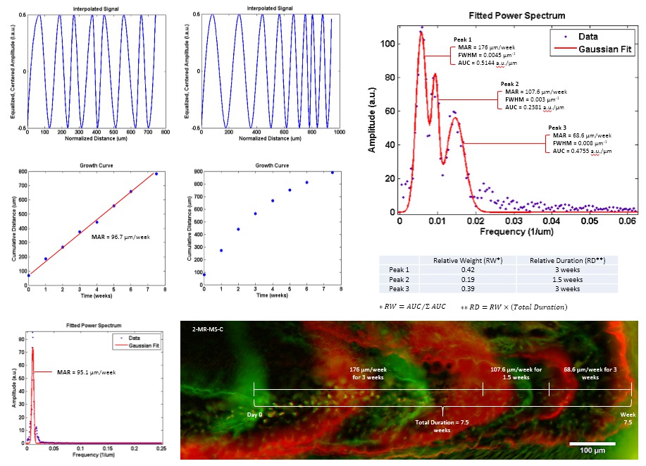
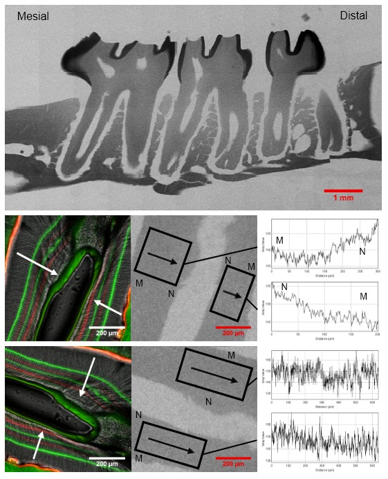
Methods: Six male, Sprague-Dawley rats (3 control/3 ligated) were administered 10 fluorochrome injections (alternating tetracycline HCL/alizarin red dyes) over 12 weeks. Hemi-maxillae were dissected, imaged, and analyzed using a Fast Fourier Transform (FFT) protocol which converts the spatial periodicity of fluorochrome labels into the frequency domain. MAR and RW was assessed in a total of 31 regions of interest (ROI’s) per specimen, and correlated with the direction of respective mineral gradients as determined via high resolution radiography. Final metrics were mapped onto corresponding ROI’s to provide a comprehensive model for understanding how periodontitis affects growth rates of dental mineralized tissues within the context of joint biomechanics.
Results: Between tissues, dentin grew the slowest but the most linear, cementum grew the fastest but the most nonlinear, and bone exhibited intermediate MAR values with site-specific nonlinear growth (Table 1). Within tissues, ligated specimens displayed accelerated distal drift where mesial portions of the maxillary complex grew significantly faster (17% in dentin, 9% in cementum, and 53% in bone; p < 0.05) than its distal portions; in controls, no such gradient was observed. The effects of periodontitis were also most pronounced in the 1st molar, where disease-related changes were primarily mediated through functionally active (mesial) sites of the maxillary dentition. Analysis of 6 specimens (93 ROI’s) was performed in ~18 hours.
Conclusions: From a biological perspective, periodontitis increases the susceptibility of dental mineralized tissues to natural distal-drift forces specifically within the mesial side of the maxillary complex. From a methods perspective, a frequency domain interpretation of MAR provides an efficient, precise, and unique mathematical approach to modeling the kinetics of mineralized tissue apposition.


