IADR Abstract Archives
Comparison of the Temporal Mandibular Joint 3D Surface and Volume Reconstructions using MRIs and CBCTs: A Pilot study.
Objectives: The aim of this study to validate a new method for reconstructing 3D models of the condyles using Magnetic Resonance Imaging (MRI).
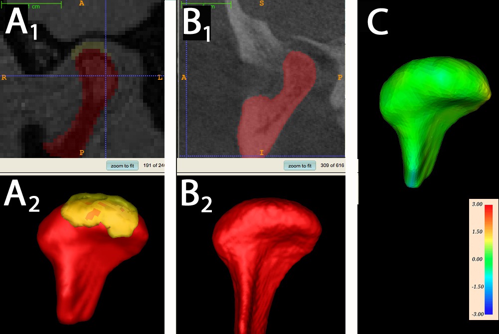
Methods: subjects with Cone Beam Computed Tomography (CBCT) and MRI taken on the same day at the University of North Carolina were enrolled in the study. A total of 10 condyles were obtained. The MRI were T2 weighted scans taken at a resolution of 0.6 x 0.6 x 0.6 mm. The CBCT were taken at a resolution of 0.5 x 0.5 x 0.5 mm. Two independent examiners built 3D models of the condyles from MRI and CBCT scans. The condylar models from the MRI and CBCT were superimposed using a procrustean best fit method. Differences between the volume and shape of the CBCT and MRI condylar models were measured using ITK-SNAP, VAM and 3D Meshmetric. Inter-examiner reliability was assessed using ICC analysis with a two-way random effect model.
Results: models built from CBCT and MRI had similar volumes (+/- 4.3 mm3) and shape (mean surface error between models: +/-0.52mm). The medial pole and posterior surface of the condylar head a showed the greatest error (mean = +/- 1.5mm). ICC analysis showed high agreement between examiners (0.939).
Conclusions: 3D models of condyles built from T2 weighted MRI scans were similar to those from CBCT scans.

