IADR Abstract Archives
Evaluation of Dentin During Caries Removal by Optical Coherence Tomography
Objectives: To evaluate the optical properties of dentin in relationship to different layers of carious dentin by optical coherence tomography (OCT), and evaluate the clinical application of OCT to assess dentin during removal of caries.
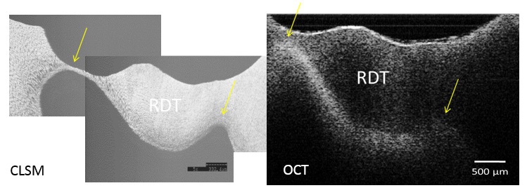
Methods: In the in vitro experiment, dentin discs including infected, affected and intact dentin were obtained from carious extracted human molars with visible occlusal cavities and scanned by a swept-source OCT at 1330nm center wavelength (Dental SS-OCT Prototype 2, Panasonic Healthcare). Attenuation coefficient (µt) and refractive index (n) were calculated for each dentin substrate. On a separate series of teeth, the remaining dentin thickness (RDT) was measured by OCT during removal of occlusal caries, and the results were compared to histological cross-sections using confocal scanning laser microscope (CLSM). In the in vivo experiment, OCT scans were obtained during excavation of occlusal caries to determine the type of dentin at the pulpal floor, and measure RDT.
Results: RDT values by OCT ranged from 140-2300µm. Pulpal horns and pulp chamber roof observation under OCT and CLSM resulted in comparable images that allowed measurement of dentin thickness; however, in the case of thick (> 2 mm) and/or caries-infected dentin tissue, the lower boundary of pulpal roof was occasionally masked due to higher µt values of infected dentin. Affected dentin showed slightly lower µt values than infected tissue. In terms of n, the following order was obtained; sound dentin>affected dentin> carious dentin. The clinical results confirmed the in vitro finding regarding the application of OCT.
Conclusions: OCT enables visualization of anatomical structures during deep caries excavation. Condition of dentin can affect OCT measurements; however, exposure of the vital dental pulp due to removal of very thin remaining dentin can be avoided with this novel non-invasive technique regardless of dentin conditions.
IMAGES
2120645_File000000.jpg
2120645_File000000.jpg