IADR Abstract Archives
Optical Imaging Demonstrates Reliable Tissue Density Quantification and Measurement Accuracy
Objectives: To establish a scale for linear measurement accuracy in OCT images and to evaluate the ability of OCT to discern between different oral tissues.
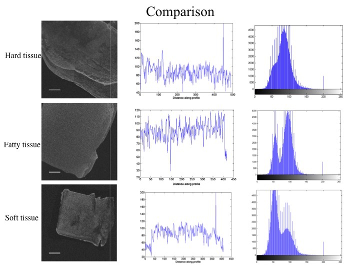
Methods: The study consisted of two phases. Phase-1 was to determine the linear measurement accuracy of OCT images using a post-processing algorithm developed in MATLAB.A 5 mm long gutta-percha point was imaged using a swept-source optical coherence tomography device (Axsun technologies Inc. Bilerica, MA, USA) operating at wavelengths ranging between 1250 nm and 1360 nm with an average power of 18 mW. Phase-2 was to image three samples resembling soft tissue (wax), fatty tissue (butter) and hard tissue (tooth enamel) with the same device. The imaging probe was placed atop a 2cm x 2cm stabilizing device to render standard imaging conditions with stable object-source distance. The images were then post-processed to evaluate the density contrasts and intensity profiles of tissues.
Results: Post processing algorithms included linear measurement along the x and y axes, histogram plots, intensity profile diagrams and contour definition that could reveal the edges and boundaries clearly. Moreover, the post-processing algorithms provided a highly accurate linear measurement of the imaged gutta-percha established by the number of pixels in the x and y axes and the resolution of the image. In the second phase of the study, significant difference was observed in the distribution of density contrast of the three images with the enamel showing the highest average density distribution followed by wax and butter. The intensity profiles however did not show any significant differences.
Conclusions: Within the limitations of this pilot study, we were able to establish a scale for linear measurement for OCT images. OCT images are able to clearly and reliably discern density contrast between different oral tissues.
IMAGES
2115341_File000000.jpg
2115341_File000000.jpg