IADR Abstract Archives
Molecular Analysis of Calcium-Phosphorous-Fluoride Based Agents in Enamel Re-Mineralization
Objectives: To assess the ability of Calcium-Phosphate delivery systems (with and without fluoride) in promoting re-mineralization of sub-surface enamel caries
To characterize the molecular structure of carious and re-mineralized enamel using Micro-Raman Spectroscopy (MRS)
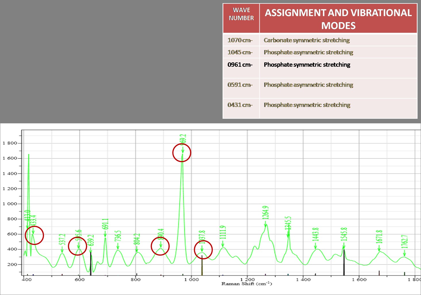
Methods: Artificial subsurface caries lesions were created on buccal surface of 108 extracted human molars. Specimens were assigned according to re-mineralizing agent into five groups:G1: Calcium Phosphate tooth paste without fluoride(Control), G2: Tri-Calcium Phosphate toothpaste (Clinpro), G3: CPP-ACP (Tooth Mousse), G4:CPP-ACP with fluoride (Tooth Mousse Plus), G5:Calcium Sodium Phosphosilicate (Novamin). Serial sections were cut through the lesions and investigated before and after 10 day pH cycling using Polarized Light Microscopy(PLM) .The differences in lesion depths obtained by PLM before and after treatment and amongst the groups was compared . A series of spectra over the cross section of lesions were taken using Micro-Raman Spectroscopy (MRS). Quantitative data on peak intensities of ν1 (960 cm-1) phosphate peak as well as B-type carbonate peak (1070 cm-1), area under the curve and width of each spectral band component were determined and mineralization was calculated. The data were statistically evaluated using one-way ANOVA for multiple comparisons.
Results: All test products showed statistically significant change in lesion depth and intensity of the ν1 (960 cm-1) phosphate peaks in the Raman spectra after re-mineralization treatments except for Control group. Clinpro showed maximum reduction in the lesion depth followed by Novamin, Tooth Mousse Plus, Tooth Mousse, with the difference being statistically insignificant. The mean of integral areas of ν1 phosphate peak among groups indicated significantly (P < 0.05) greater mineralization in the Clinpro and Novamin group.
Conclusions: Newer dentifrices based on innovative technology, showed good re-mineralization potential by making more Calcium-Phosphate-Fluoride ions available at site of re-mineralization. Raman spectroscopy is useful tool for analyzing molecular structure of healthy and re-mineralized enamel.
To characterize the molecular structure of carious and re-mineralized enamel using Micro-Raman Spectroscopy (MRS)
Methods: Artificial subsurface caries lesions were created on buccal surface of 108 extracted human molars. Specimens were assigned according to re-mineralizing agent into five groups:G1: Calcium Phosphate tooth paste without fluoride(Control), G2: Tri-Calcium Phosphate toothpaste (Clinpro), G3: CPP-ACP (Tooth Mousse), G4:CPP-ACP with fluoride (Tooth Mousse Plus), G5:Calcium Sodium Phosphosilicate (Novamin). Serial sections were cut through the lesions and investigated before and after 10 day pH cycling using Polarized Light Microscopy(PLM) .The differences in lesion depths obtained by PLM before and after treatment and amongst the groups was compared . A series of spectra over the cross section of lesions were taken using Micro-Raman Spectroscopy (MRS). Quantitative data on peak intensities of ν1 (960 cm-1) phosphate peak as well as B-type carbonate peak (1070 cm-1), area under the curve and width of each spectral band component were determined and mineralization was calculated. The data were statistically evaluated using one-way ANOVA for multiple comparisons.
Results: All test products showed statistically significant change in lesion depth and intensity of the ν1 (960 cm-1) phosphate peaks in the Raman spectra after re-mineralization treatments except for Control group. Clinpro showed maximum reduction in the lesion depth followed by Novamin, Tooth Mousse Plus, Tooth Mousse, with the difference being statistically insignificant. The mean of integral areas of ν1 phosphate peak among groups indicated significantly (P < 0.05) greater mineralization in the Clinpro and Novamin group.
Conclusions: Newer dentifrices based on innovative technology, showed good re-mineralization potential by making more Calcium-Phosphate-Fluoride ions available at site of re-mineralization. Raman spectroscopy is useful tool for analyzing molecular structure of healthy and re-mineralized enamel.
IMAGES
2107454_File000000.jpg
2107454_File000000.jpg