IADR Abstract Archives
Periapical Lesions – The Need to Biopsy?
Objectives: The purpose of this study is to examine and compare clinical/radiographic impressions and histological diagnoses of specimens clinically believed to be periapical granulomas, radicular cysts, or other periapical pathoses. Specifically, estimate the prevalence of malignancies in oral pathology specimens thought to be one of the above mentioned benign lesions.
Methods:
Records were extracted from the VCU Oral Pathology biopsy service database containing 168,095 cases biopsied from late 1987 through April 2011. Specific variables of interest queried included contributors clinical impression, final diagnosis of the oral pathologist, location of specimen, biopsy data, contributor’s practice type, patient’s sex, DOB and age. The data sets included records for which the submitting clinical impression was periapical granuloma, radicular cyst, or periapical pathosis, and included patients in the age range of 10 to 100 years.
Results:
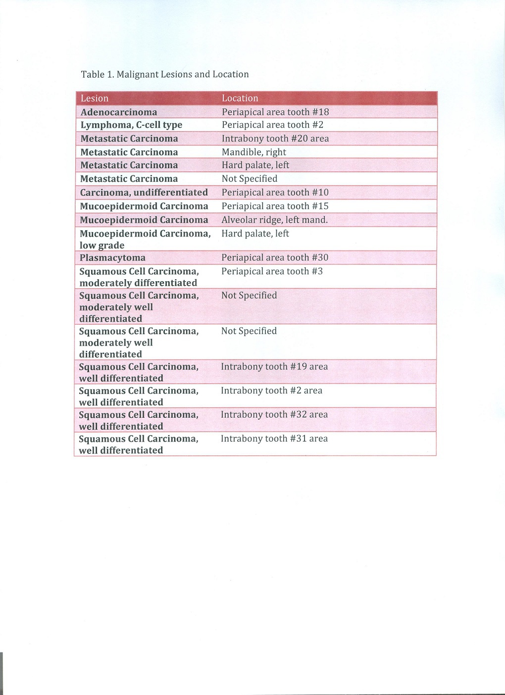
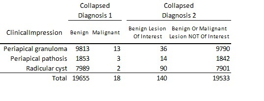
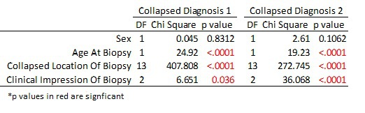
The data set contained 19,673 patient records. The clinical/radiographic impressions of the biopsy specimens at the time of submission were Periapical Granuloma 9826 (50%), Periapical Pathosis (9%), and Radicular Cyst 7991 (41%). Diagnoses revealed several unique conditions of which 18 were malignancies including: adenocarcinoma, lymphoma, metastatic carcinoma, mucoepidermoid carcinoma, squamous cell carcinoma and plasmacytoma. Bivariate analysis of possible predictor variables found Age of Biopsy, Location and Clinical Impression to be significant (p<0.05).
Conclusions:
While the number of malignant lesions in this large series of biopsies is extremely small, it nonetheless demonstrates that a clinician cannot assume the nature of the excised lesion to be benign and therefore choose not to submit the specimen for histological analysis.
Methods:
Records were extracted from the VCU Oral Pathology biopsy service database containing 168,095 cases biopsied from late 1987 through April 2011. Specific variables of interest queried included contributors clinical impression, final diagnosis of the oral pathologist, location of specimen, biopsy data, contributor’s practice type, patient’s sex, DOB and age. The data sets included records for which the submitting clinical impression was periapical granuloma, radicular cyst, or periapical pathosis, and included patients in the age range of 10 to 100 years.
Results:
The data set contained 19,673 patient records. The clinical/radiographic impressions of the biopsy specimens at the time of submission were Periapical Granuloma 9826 (50%), Periapical Pathosis (9%), and Radicular Cyst 7991 (41%). Diagnoses revealed several unique conditions of which 18 were malignancies including: adenocarcinoma, lymphoma, metastatic carcinoma, mucoepidermoid carcinoma, squamous cell carcinoma and plasmacytoma. Bivariate analysis of possible predictor variables found Age of Biopsy, Location and Clinical Impression to be significant (p<0.05).
Conclusions:
While the number of malignant lesions in this large series of biopsies is extremely small, it nonetheless demonstrates that a clinician cannot assume the nature of the excised lesion to be benign and therefore choose not to submit the specimen for histological analysis.
IMAGES
2099502_File000000.jpg
2099502_File000001.jpg
2099502_File000002.jpg
2099502_File000000.jpg
2099502_File000001.jpg
2099502_File000002.jpg