IADR Abstract Archives
Impacted Maxillary Canine: 3-D Assessment of Localization and Severity
Objectives: To determine the location, position and severity of impacted or unerupted maxillary canines as they correlate to clinical aspects of orthodontics. To identify specific regions in the maxilla where they are more common. To evaluate these methods for reliability and accuracy. To introduce a novel 3-D classification for maxillary impacted canines.
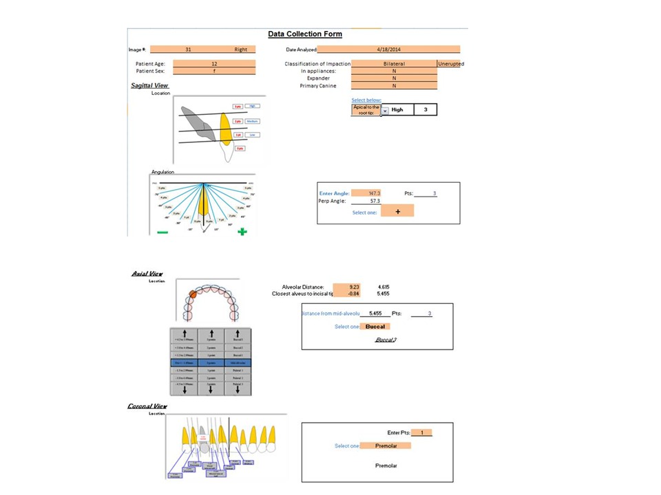
Methods: Out of 1000 CBCT images of patients with impacted maxillary canines, 207 CBCTs, with 314 unerupted canines were selected. Canine was classified as impacted based on either complete root development or erupted contralateral canine. Of 314 unerupted canines,174 were impacted.
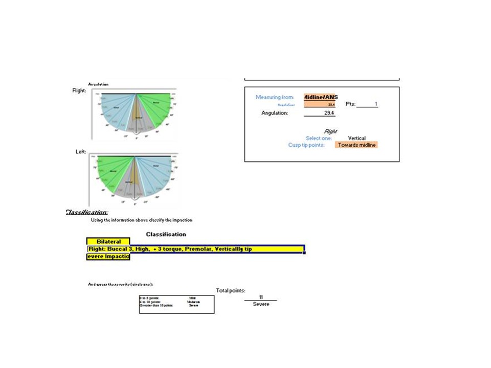
Results: Excellent inter and intra-examiner reliability was obtained for all variables except axial deviations from the midline. Measurements comparing those from a digital caliper and CBCT image on a typodont showed high similarity. Overall, females were more commonly effected; 1.63:1. It increased to 1.93:1 when impacted. Rates of palatal, buccal and midalveolar canine locations were 38.54%, 40.76% and 20.70% respectively. When impacted, palatal to buccal displacement was 2.14:1. Coronally, 34.08% were normally positioned. Overall, 62.10% were mesial to the distal border of the lateral incisor and when impacted it increased to 78.19%. For canines located mesial to the midline of the lateral incisor the overall and impacted values were 45.54% and 60.92%. Rate of mesially tipped canines was 59.24% for overall and 78.29% when impacted. Canines were also classified as mild 33.44%, moderate 37.58% and severe28.98% based on the findings. Yearly increase in age, showed 3.2% increased chance of severe impaction.
Conclusions: Location of impacted / unerupted maxillary canines was evaluated by position, angulation and severity. Specific regions on the maxilla were identified as common locations. There was a positive correlation between age and severity. The methods were found to be reliable and accurate. A classification for impacted maxillary canines examined by CBCT imaging was introduced.
Methods: Out of 1000 CBCT images of patients with impacted maxillary canines, 207 CBCTs, with 314 unerupted canines were selected. Canine was classified as impacted based on either complete root development or erupted contralateral canine. Of 314 unerupted canines,174 were impacted.
Results: Excellent inter and intra-examiner reliability was obtained for all variables except axial deviations from the midline. Measurements comparing those from a digital caliper and CBCT image on a typodont showed high similarity. Overall, females were more commonly effected; 1.63:1. It increased to 1.93:1 when impacted. Rates of palatal, buccal and midalveolar canine locations were 38.54%, 40.76% and 20.70% respectively. When impacted, palatal to buccal displacement was 2.14:1. Coronally, 34.08% were normally positioned. Overall, 62.10% were mesial to the distal border of the lateral incisor and when impacted it increased to 78.19%. For canines located mesial to the midline of the lateral incisor the overall and impacted values were 45.54% and 60.92%. Rate of mesially tipped canines was 59.24% for overall and 78.29% when impacted. Canines were also classified as mild 33.44%, moderate 37.58% and severe28.98% based on the findings. Yearly increase in age, showed 3.2% increased chance of severe impaction.
Conclusions: Location of impacted / unerupted maxillary canines was evaluated by position, angulation and severity. Specific regions on the maxilla were identified as common locations. There was a positive correlation between age and severity. The methods were found to be reliable and accurate. A classification for impacted maxillary canines examined by CBCT imaging was introduced.
IMAGES
2097602_File000000.jpg
2097602_File000001.jpg
2097602_File000000.jpg
2097602_File000001.jpg