IADR Abstract Archives
N-methylene Phosphonic Chitosan – A Novel Induced Template for Enamel Biomineralization
Objectives: Modified phosphoric chitosan molecules model to induce the crystallization of hydroxyapatite with 3D-structure in a controllable way which realize the biomineralization of enamel in vitro.
Methods: N-methylene phosphonic chitosan (NPCS) was synthesized via formal dehyde addition and condensation with phosphoric acid in a step reaction. Human molars were sliced into 1mm thickness disks and etched with 0.5N EDTA to reveal the different orientations of the dentinal tubule. The molars disks were daubed with phosphate ions dental adhesive agent equably (Prime& BondNT, DentsplyDetreyGmbh). The solidification process induced by curing light is 10 s at least. N-methylene phosphonic chitosan solution(test group) and chitosan solution(contrast group) were daubed to disks of molar after phosphorylation. CaCl2 and Na3PO4-12H2O solutions were added after the crosslinking process. The obtained composite were characterized by Fourier transformed infrared spectroscopy (FTIR) and X-ray diffraction (XRD) as well as scanning electron microscopy (SEM).
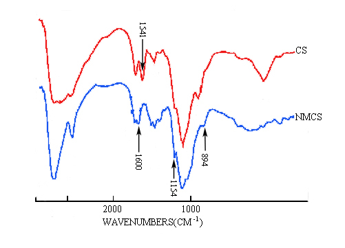
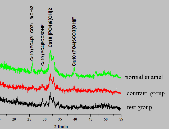
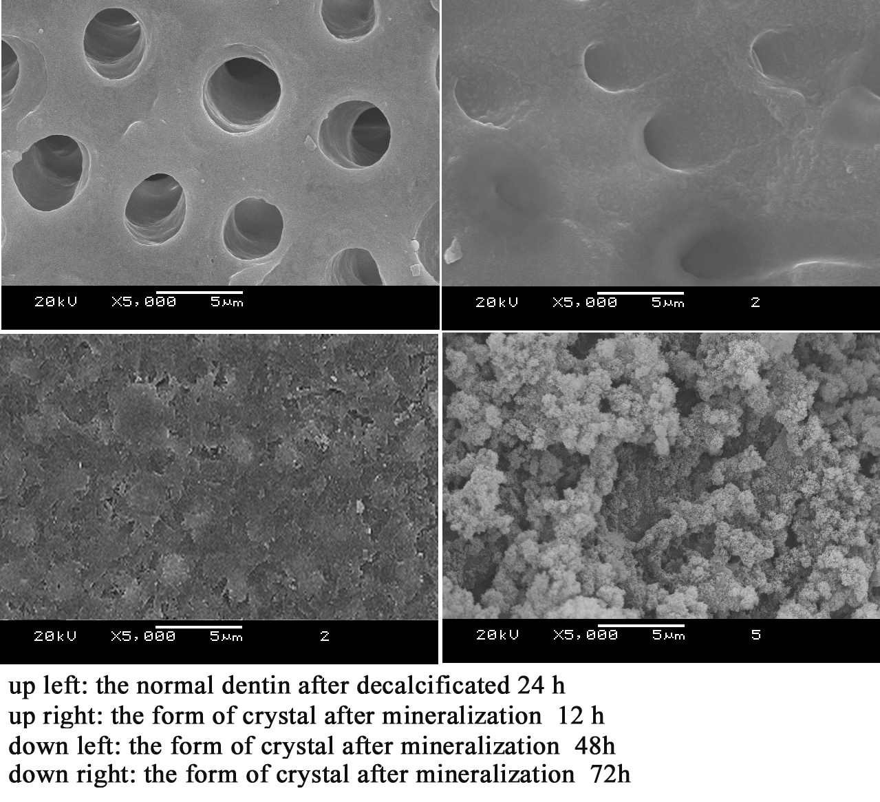
Results: N-methylene phosphonic chitosan could induce the crystallization in solutions and crosslink to phosphate ions remained on surface of dentin. FTIR spectrogram(Figure 1) showed there are absoprtion band of chitosan (894 cm-1 ) and N-methylene phosphonic chitosan (1154 cm-1) on the surface of molar disks, which were the characteristic band of glycosyl in chitosan. The organic compounds construct the three dimensional frame during nucleation and growth stage of hydroxyapatite which confirmed by XRD and SEM. XRD spectrum of the neonatal crystal (Figure 2) suggested that several diffraction peaks around 2θ=32˚ which are corresponding to the expected Bragg peaks for hydroxyapatite, implying that HA crystals were formed. The dentinal tubule were blocked by neonatal hydroxyapatite layer with a continuous structure of columns which size between 10-20nm(Figure 3).
Conclusions: The results showed that phosphonic chitosan monolayer can be used as a potential effective modifier in inchoate enamel remineralization.
Methods: N-methylene phosphonic chitosan (NPCS) was synthesized via formal dehyde addition and condensation with phosphoric acid in a step reaction. Human molars were sliced into 1mm thickness disks and etched with 0.5N EDTA to reveal the different orientations of the dentinal tubule. The molars disks were daubed with phosphate ions dental adhesive agent equably (Prime& BondNT, DentsplyDetreyGmbh). The solidification process induced by curing light is 10 s at least. N-methylene phosphonic chitosan solution(test group) and chitosan solution(contrast group) were daubed to disks of molar after phosphorylation. CaCl2 and Na3PO4-12H2O solutions were added after the crosslinking process. The obtained composite were characterized by Fourier transformed infrared spectroscopy (FTIR) and X-ray diffraction (XRD) as well as scanning electron microscopy (SEM).
Results: N-methylene phosphonic chitosan could induce the crystallization in solutions and crosslink to phosphate ions remained on surface of dentin. FTIR spectrogram(Figure 1) showed there are absoprtion band of chitosan (894 cm-1 ) and N-methylene phosphonic chitosan (1154 cm-1) on the surface of molar disks, which were the characteristic band of glycosyl in chitosan. The organic compounds construct the three dimensional frame during nucleation and growth stage of hydroxyapatite which confirmed by XRD and SEM. XRD spectrum of the neonatal crystal (Figure 2) suggested that several diffraction peaks around 2θ=32˚ which are corresponding to the expected Bragg peaks for hydroxyapatite, implying that HA crystals were formed. The dentinal tubule were blocked by neonatal hydroxyapatite layer with a continuous structure of columns which size between 10-20nm(Figure 3).
Conclusions: The results showed that phosphonic chitosan monolayer can be used as a potential effective modifier in inchoate enamel remineralization.
IMAGES
2093579_File000000.jpg
2093579_File000001.jpg
2093579_File000002.jpg
2093579_File000000.jpg
2093579_File000001.jpg
2093579_File000002.jpg