IADR Abstract Archives
THE MANDIBULAR INCISIVE CANAL: VISIBILITY AND LOCATION ON CONE-BEAM COMPUTED TOMOGRAPHY IMAGES IN A GROUP OF THAI
Objectives: To assess the visibility, location and size of the mandibular incisive canal (MIC) in a group of Thai by using cone-beam computed tomography (CBCT) images.
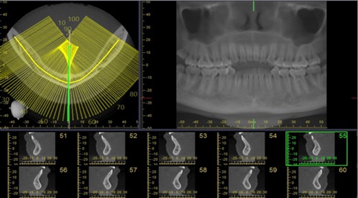
Methods: Retrospective study of 120 CBCT images in the anterior mandible were performed. All CBCT images were obtained with 3D Accuitomo (J. Morita Mfg. Corp., Kyoto, Japan) with the exposure setting of 80-90 kVp, 4-5 mA, 18 s., and FOV 10×10 cm with voxel size of 0.25 mm. The assessment of the visibility of the MIC will be recorded the detection frequencies and analyzed into the percentage. For linear measurement, the CBCT images will be analyzed from the curvedMPR. The cross-sectional images that pass through the midline, 5 mm, 10 mm, 15 mm, 20 mm from the midline (both right and left sides) will be selected and measured with the digital ruler from the viewer (i-dixel) equipped with the CBCT machine. All images will be measured in the following distances: Incisive canal to the inferior border of mandible (L1), Incisive canal to buccal plate of mandible (L2), Incisive canal to lingual plate of mandible (L3), Bucco-lingual diameter of outer contour of MIC (D1), Vertical diameter of outer contour of MIC (D2).
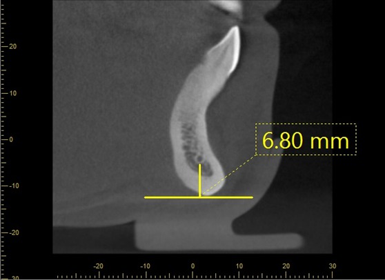
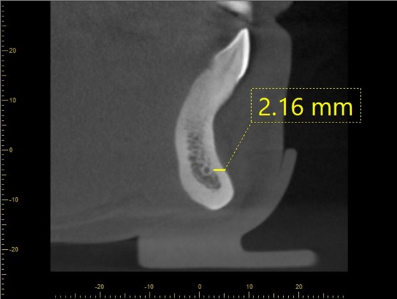
Results: The MIC was identified in 96% of the CBCT images. Mean (s.d.) distance from MIC to inferior border of mandible at midline, 5 mm, 10 mm, 15 mm, 20 mm from the midline were 8.74 (1.61) mm, 9.80 (1.37) mm, 10.21 (1.12) mm, 10.13 (1.89) mm, 11.03 (1.56) mm, respectively. Mean (s.d.) distance from MIC to buccal border of mandible were 4.17 (1.41) mm, 3.86 (1.28) mm, 3.74 (1.19) mm, 3.24 (1.03) mm, 3.18 (1.31) mm, respectively. Mean (s.d.) distance from MIC to lingual border of mandible were 4.02 (1.17) mm, 3.46 (1.06) mm, 3.54 (1.62) mm, 3.29 (1.47) mm, 3.84 (1.71) mm, respectively. Mean (s.d.) buccolingual diameter were 1.78 (0.37) mm, 1.97 (0.52) mm, 2.10 (0.40) mm, 2.25 (0.73) mm, 2.86 (0.51) mm, respectively. Mean (s.d.) vertical diameter were 1.84 (0.37) mm, 1.91 (0.49) mm, 2.53 (0.38) mm, 2.92 (0.46) mm, 3.11 (0.43) mm, respectively.
Conclusions: A well-defined MIC could be identified in the majority of the CBCT images. The diameter of the MIC is rather large that might have an important clinical implication in relation to surgery. CBCT demonstrates to be a good presurgical assessment before the surgical procedure in the anterior mandible.
Methods: Retrospective study of 120 CBCT images in the anterior mandible were performed. All CBCT images were obtained with 3D Accuitomo (J. Morita Mfg. Corp., Kyoto, Japan) with the exposure setting of 80-90 kVp, 4-5 mA, 18 s., and FOV 10×10 cm with voxel size of 0.25 mm. The assessment of the visibility of the MIC will be recorded the detection frequencies and analyzed into the percentage. For linear measurement, the CBCT images will be analyzed from the curvedMPR. The cross-sectional images that pass through the midline, 5 mm, 10 mm, 15 mm, 20 mm from the midline (both right and left sides) will be selected and measured with the digital ruler from the viewer (i-dixel) equipped with the CBCT machine. All images will be measured in the following distances: Incisive canal to the inferior border of mandible (L1), Incisive canal to buccal plate of mandible (L2), Incisive canal to lingual plate of mandible (L3), Bucco-lingual diameter of outer contour of MIC (D1), Vertical diameter of outer contour of MIC (D2).
Results: The MIC was identified in 96% of the CBCT images. Mean (s.d.) distance from MIC to inferior border of mandible at midline, 5 mm, 10 mm, 15 mm, 20 mm from the midline were 8.74 (1.61) mm, 9.80 (1.37) mm, 10.21 (1.12) mm, 10.13 (1.89) mm, 11.03 (1.56) mm, respectively. Mean (s.d.) distance from MIC to buccal border of mandible were 4.17 (1.41) mm, 3.86 (1.28) mm, 3.74 (1.19) mm, 3.24 (1.03) mm, 3.18 (1.31) mm, respectively. Mean (s.d.) distance from MIC to lingual border of mandible were 4.02 (1.17) mm, 3.46 (1.06) mm, 3.54 (1.62) mm, 3.29 (1.47) mm, 3.84 (1.71) mm, respectively. Mean (s.d.) buccolingual diameter were 1.78 (0.37) mm, 1.97 (0.52) mm, 2.10 (0.40) mm, 2.25 (0.73) mm, 2.86 (0.51) mm, respectively. Mean (s.d.) vertical diameter were 1.84 (0.37) mm, 1.91 (0.49) mm, 2.53 (0.38) mm, 2.92 (0.46) mm, 3.11 (0.43) mm, respectively.
Conclusions: A well-defined MIC could be identified in the majority of the CBCT images. The diameter of the MIC is rather large that might have an important clinical implication in relation to surgery. CBCT demonstrates to be a good presurgical assessment before the surgical procedure in the anterior mandible.
IMAGES
2081216_File000000.jpg
2081216_File000001.jpg
2081216_File000002.jpg
2081216_File000000.jpg
2081216_File000001.jpg
2081216_File000002.jpg